Domain 10 — Synthesis & Recommendations (Deepened)¶
⚠️ MEDICAL DISCLAIMER: This document is a research synthesis compiled from peer-reviewed literature, clinical guidelines, public medical information, and raw research sources. It is NOT medical advice, a treatment plan, or a substitute for professional medical judgment. All recommendations must be reviewed and individualized by the patient's treating medical team. Decisions about diagnosis, treatment, and surgical intervention must be made in consultation with qualified healthcare professionals. The evidence levels cited reflect published literature quality, not certainty of individual outcomes.
Status: Deepened Research Synthesis & Integrated Recommendations Clinical Focus: Adult post-surgical clubfoot complications — chronic wounds, progressive deformity, and mobility loss following childhood corrective surgery Last Updated: 2026-05-15 Last Reviewed: 2026-05-16 | ⚠️ Evidence Grading Note: Evidence levels in this document reflect the best available literature for adult post-clubfoot surgical complications. Many recommendations are supported by Level IV evidence (case series, retrospective cohorts) rather than Level I-II, because no RCTs exist for this population. The evidence grades in the tables below should be read as "best available evidence" rather than "confidence level." Where Level I-II tags appear, they reflect the type of source (systematic review, prospective study) rather than strong recommendation strength. Clinical decision-makers should apply GRADE methodology to determine recommendation strength for this specific patient. | ⚠️ Clinical Sequencing Note: This document presents treatment options as a sequential clinical pathway, not a parallel menu of equivalents. The intended reading order is: Diagnose → Stabilize → Treat → Reconstruct → Rehabilitate. See the Prioritized Action Plan (Section 1) for the correct clinical sequence. Do not treat the treatment options sections as equivalent alternatives to be considered simultaneously.
SEQUENTIAL CLINICAL PATHWAY — READ IN THIS ORDER¶
⚠️ This document is structured as a sequential clinical journey, not a parallel menu. Each phase builds on the previous. Do not skip ahead. Follow this pathway:
| Step | Clinical Phase | Document Sections | Key Decision Point |
|---|---|---|---|
| 1 | Diagnose | §1.2 Diagnostic Workup, §6.2-6.3 Pathophysiology, §7 Decision Frameworks | What is the current state of the foot? Vascular? Neurological? Structural? |
| 2 | Optimize | §1.1 Team Assembly, §1 PHASE 1-2, §4 Red Flags, §6.4 Pre-Surgical Workup | Clear infection, optimize wound bed, medical clearance, bone density, psychological readiness |
| 3 | Decide | §2 Treatment Pathways (compare ONLY after Steps 1-2), §7 Decision Frameworks, §8 Nonunion Risk | Which pathway fits the affected adult's? Triple arthrodesis vs TSF vs amputation? |
| 4 | Treat | §2 Primary/Alternative Pathways, §9 Limb Salvage Protocol | Execute the chosen surgical pathway with full team coordination |
| 5 | Rehabilitate | §1 PHASE 4, §6 Rehabilitation domain, §10 Cost/Insurance | Long-term recovery, offloading, prevention of recurrence |
| 6 | Contingency | §1 PHASE 5, §7.2 Salvage vs Amputation Algorithm | Only if Steps 1-5 fail — LEAP-informed amputation decision |
The specialist consultation checklist (§3) and patient-facing summary (§5) are reference materials to use WITHIN each step, not standalone sections to read independently.
TABLE OF CONTENTS¶
- PRIORITIZED ACTION PLAN — Start here. The sequential clinical pathway.
- TREATMENT PATHWAY COMPARISON — Compare pathways AFTER completing Phase 1-2 optimization
- SPECIALIST CONSULTATION CHECKLIST — Questions to bring to each specialist at each step
- RED FLAGS & CONTRAINDICATIONS — Must be cleared BEFORE proceeding to surgical decision (Step 2)
- PATIENT-FACING SUMMARY — Share with patient at initial consultation
- MEDICAL PROFESSIONAL SUMMARY — Share with referring physicians
- DECISION FRAMEWORK — Use during Step 3 (Decide) to choose the right pathway
- NONUNION RISK FACTORS — Factor into Step 3 risk-benefit analysis
- MULTIDISCIPLINARY LIMB SALVAGE PROTOCOL — Execution protocol for Step 4 (Treat)
- COST AND INSURANCE CONSIDERATIONS — Pre-authorize before Step 4
Cross-links: [[index]] | [[domain-1-root-cause-surgical-complications]] | [[domain-2-diagnosis]] | [[domain-3-wound-care]] | [[domain-4-surgical-correction]] | [[domain-5-vascular-neurological]] | [[domain-6-rehabilitation]] | [[domain-7-emerging]] | [[domain-8-specialists-centers]] | [[domain-9-medical-legal]]
1. PRIORITIZED ACTION PLAN¶
Synthesized from all nine domains. Ordered from most immediate to longest-term actions. Each phase assumes the prior phase's goals have been substantially met.
PHASE 1: IMMEDIATE (Weeks 1–4) — Stabilize & Assess¶
1.1 Assemble Multidisciplinary Team¶
Priority: Critical
Per [[domain-8-specialists-centers]], the minimum care team must include:
| # | Specialist | Why | First Appointment Focus | Evidence Level |
|---|---|---|---|---|
| 1 | Orthopaedic Foot & Ankle Surgeon (fellowship-trained) | Primary architect of reconstruction plan | Initial deformity assessment, pattern classification ([Domain 1]) | V (expert consensus) |
| 2 | Vascular Surgeon | Assess arterial inflow before any surgery | ABI, duplex ultrasound, angiography if ABI<0.9 | I (Conte 2019 Global Vascular Guidelines) |
| 3 | Wound Care Specialist | Stabilize chronic wounds | Wound classification (Wagner/UT), infection workup, NPWT consideration | I (IWGDF 2023) |
| 4 | Physiatrist (PM&R) | Gait analysis, offloading strategy | Baseline mobility, AFO candidacy, pain management | III (Dobbs 2006) |
| 5 | Plastic/Reconstructive Surgeon | Soft tissue coverage if needed | Flap planning for compromised envelope | III (Fijany 2023) |
How to select the first specialist (criteria-first): Geography is a filter, not a ranking. Look for an orthopaedic foot & ankle surgeon who is fellowship-trained in complex adult deformity / revision clubfoot, practices in a setting with access to vascular surgery, wound care, and plastic/reconstructive coverage, and has documented high-volume complex foot reconstruction or limb-salvage experience (Level V — selection criteria; see [[domain-8-specialists-centers]] and [[top-tier-specialist-directory]]). Use AOFAS Find-a-Surgeon and institutional limb-salvage program pages to verify credentials before contact.
Example centers meeting these criteria (peers by capability; region is an attribute only — verify current contact info before calling):
| Example center | Region | Capability notes |
|---|---|---|
| Hospital for Special Surgery (HSS) Foot & Ankle | New York, NY | High-volume foot & ankle; adult residual-clubfoot 3D assessment work presented internationally |
| Mayo Clinic | Rochester, MN | Full-spectrum orthopaedics + regenerative / limb programs |
| Cleveland Clinic | Cleveland, OH | Integrated vascular + ortho + plastic + wound care limb salvage |
| UW Medicine / Harborview Foot & Ankle | Seattle, WA | Level I trauma / complex deformity and limb-salvage programs |
| OHSU Foot & Ankle | Portland, OR | Fellowship-trained adult clubfoot focus within multidisciplinary limb preservation |
Contact path and last-verified details: [[top-tier-specialist-directory]].
1.2 Complete Diagnostic Workup¶
Priority: CriticalImaging ([domain-2-diagnosis]): - Weight-bearing bilateral X-rays (AP, lateral, oblique) — current deformity and joint status (Level III) - CT with 3D reconstruction — bone architecture, prior surgical alterations, planning for arthrodesis — essential given that standard X-rays underestimate post-PMR 3D deformity (Level IV, [[domain-1-root-cause-surgical-complications]]) - MRI of feet/ankles — soft tissue scarring, tendon integrity, osteomyelitis screening (Level IV)
Vascular Studies ([domain-5-vascular-neurological]): - Ankle-Brachial Index (ABI) bilateral — perfusion baseline (Level I) - Arterial duplex ultrasound — posterior tibial and dorsalis pedis patency (Level I) - Transcutaneous oxygen tension (TcPO2) if ABI abnormal — threshold for healing >30 mmHg (Level I, Conte 2019) - Toe-Brachial Index (TBI) — microvascular assessment, unaffected by calcification (Level IV)
Neurological Evaluation ([domain-5-vascular-neurological]): - Nerve conduction studies / EMG — tarsal tunnel, peripheral neuropathy assessment (Level III-IV, Dellon 1999 (PMID 10563271)) - Semmes-Weinstein monofilament testing — protective sensation (Level I) - Tinel's sign at medial malleolus and fibular neck (Level III)
Wound-Specific ([domain-3-wound-care]): - Wound culture and bone biopsy if osteomyelitis suspected (Level I, IWGDF) - Plantar pressure mapping (pedobarography) — identify pressure points driving wound formation ([domain-1-root-cause-surgical-complications] deformity → wound location table) - UT WIfI classification — standardized wound, ischemia, foot infection grading (Level IIb, Mills 2014)
Laboratory: - ESR, CRP — inflammatory markers for osteomyelitis screening (Level IV) - HbA1c — rule out undiagnosed diabetes as confounder (Level I) - Complete blood count — baseline, infection screening (Level I)
1.3 Attempt Medical Records Retrieval¶
Priority: High
Contact hospitals where childhood surgery was performed. Request operative reports, pre/post-op X-rays, anesthesia records. For records from the 1970s–1980s, expect many may have been destroyed — WA requires only ~26 years retention; OR requires 10 years from last contact ([domain-9-medical-legal]). Alternative: experienced surgeons can reverse-engineer surgical history from current CT/MRI.
If retrievable, specifically seek: - Which PMR technique was used (Turco vs. McKay vs. other) - Which tendons were lengthened or divided - Number of prior surgeries (single vs. multiple — Dobbs found 78.4 vs 64.8 functional score, p<0.005) - Age at each surgery - Any documented complications
PHASE 2: SHORT-TERM (Months 1–3) — Optimize Wound Bed & Medical Clearance¶
2.1 Wound Optimization Protocol ([domain-3-wound-care])¶
First-line — Offloading (the single most critical intervention): - Total Contact Casting (TCC) — reduces plantar pressure by 80–90% vs standard footwear; 70–90% complete healing at 12–20 weeks (Level I for diabetic ulcers per IWGDF; Level IV extrapolated for non-diabetic structural wounds) - Custom-molded TCC (not standard) — required for post-clubfoot anatomy with irregular bony prominences (Level III, Farha 2021) - Irremovable walker boot — alternative if TCC contraindicated; ~70–75% pressure reduction (Level IV)
Second-line — Advanced Wound Therapies ([domain-7-emerging]): - NPWT/VAC — Level I for diabetic foot ulcers (not validated for non-diabetic structural wounds); accelerates healing ~30–50% vs standard care; bridge therapy for larger wounds (Level IV for this population) - PRP injections — 2025 meta-analyses show ~75% complete healing vs ~50% standard care; autologous, low-risk (Level III-IV, small heterogeneous trials) - Bioengineered skin substitutes (Apligraf, Dermagraft) — 22+ RCTs supporting use when standard care fails (Level I for diabetic ulcers; Level IV for non-diabetic structural wounds)
Third-line — Adjunctive: - HBOT — mixed evidence; CMS coverage limited; may help if TcPO2 <30 mmHg documented (Level IIb, conflicting) - Stem cell therapy — emerging; effective per 2025 umbrella review (Level IV-V, no RCTs in non-diabetic structural wounds)
If osteomyelitis present ([domain-4-surgical-correction]): - Bone biopsy and culture-directed antibiotics - Surgical debridement mandatory before reconstructive planning - Hindfoot osteomyelitis carries 50% risk of above-ankle amputation (Level IV, clinical literature; specific source not traceable) - Coordinate timing with infectious disease specialist
2.2 Pain Management and Gait Preservation¶
- Custom offloading AFOs — reduce abnormal plantar pressure on both sides ([domain-6-rehabilitation])
- Custom orthotics with pressure redistribution for bilateral compensation
- Physical therapy evaluation — maintain mobility, prevent secondary contractures
- Pain management referral — neuropathic pain may require gabapentin/pregabalin if tarsal tunnel confirmed ([domain-5-vascular-neurological])
2.3 Pre-Surgical Medical Optimization¶
- Vascular optimization: If ABI <0.7, consider endovascular angioplasty/stenting first (Level Ib, BASIL-2 trial, Bradbury et al. 2023)
- Nerve decompression evaluation: If NCS confirms tarsal tunnel syndrome, decompression may restore protective sensation before wound closure (Level III-IV, Dellon 1999 (PMID 10563271))
- Smoking cessation if applicable — nonunion risk dramatically elevated in arthrodesis (see Section 8)
- Nutritional optimization — albumin >3.5 g/dL, prealbumin >20 mg/dL for wound healing (Level III)
- Bone density screening (DEXA scan) — midlife adults with chronic mobility limitation are at elevated risk for osteopenia/osteoporosis, which directly impacts arthrodesis healing rates and implant fixation. DEXA should be obtained pre-operatively if the patient has been non-weight-bearing or limited-weight-bearing for extended periods. If T-score <−1.5, consider bisphosphonate optimization or vitamin D/calcium supplementation before fusion surgery (Level IV, expert consensus — no RCTs in this specific population)
- Psychological assessment and readiness — chronic pain, progressive disability, and the prospect of major reconstructive surgery or amputation carry significant psychological burden. Screening for depression (PHQ-9), anxiety (GAD-7), and pain catastrophizing (PCS) should be performed. Psychological readiness affects surgical compliance, rehabilitation engagement, and outcomes. Consider referral to health psychologist or pain psychologist pre-operatively (Level IV, expert consensus).
- Medication review — discontinue anticoagulants/antiplatelets per surgeon protocol; assess for medications that impair bone healing (chronic corticosteroids, certain antidepressants, PPIs at high dose)
PHASE 3: MEDIUM-TERM (Months 3–9) — Surgical Reconstruction¶
3.1 Surgical Decision Point¶
Based on completed diagnostic workup and wound optimization:
| Condition | Action |
|---|---|
| Wounds healed or controllable AND ABI >0.7 | → Proceed to surgical reconstruction planning |
| Wounds actively infected or osteomyelitis documented | → Address infection first (debridement, antibiotics). Reconstruction deferred |
| ABI <0.4, non-revascularizable | → Limb salvage may not be viable. Amputation discussion warranted |
| BMI >30 | → Strong negative correlation (r=−0.738) with arthrodesis outcomes. Consider weight optimization first |
| Bilateral disease | → Sequential surgery (6-month interval) preserves mobility on one side |
3.2 Primary Surgical Pathway: Triple Arthrodesis¶
Confidence: High — strongest evidence base for post-clubfoot salvage [[domain-4-surgical-correction]]
- 81–100% patient satisfaction in clubfoot populations (Level III-IV, retrospective case series; Zhuang 2019; Maier 2023)
- AOFAS improvement: +25–43 points (Level IV, case series; Vietnam 2022: 40.6→83.8, p<0.01)
- Creates a stable, plantigrade foot — directly addresses the structural root cause of wound formation
- Peak salvage surgery age: 55–74 years — adult patients in this demographic is entering the surgical window
- Recovery: 6–8 weeks NWB, mean 69 outpatient PT sessions, 9.6 months return to work (range 3–34.5mo) (Level IV, retrospective case series; Maier 2023)
If ankle arthritis also present (57–67% of triple arthrodesis patients develop it): → Consider combined triple arthrodesis + ankle arthrodesis (Level IV, retrospective cohort; Zhuang 2019)
If patient is highly active and soft tissue permits: → Total Ankle Arthroplasty (Fijany 2023 case) — preserves motion but requires intact soft-tissue envelope and no active infection. Currently contraindicated if chronic wounds are present (Level IV)
3.3 Alternative/Adjunctive Surgical Options¶
| Option | When Appropriate | Evidence | Evidence Level |
|---|---|---|---|
| Ilizarov/TSF external fixation | Severe multiplanar deformity, gradual correction needed, joint preservation desired | 81.4% pooled success rate; 19.6–27% pin tract infection | IV (Vaccalluzzo et al. 2025 meta-analysis of case series) |
| Closing wedge osteotomy | Milder, flexible deformity | AOFAS improvement of +42.9 points; 100% plantigrade | IV (retrospective case series) |
| Tendon transfer (ATT→PT) | Muscle imbalance after bony alignment corrected | 1.43% complication rate; n=176, all angles improved p<0.001 | IIb (2024 study) |
| Staged reconstruction | Worst cases: TSF → arthrodesis → free flap | Pain-free, normal shoes at 1yr | IV (Kadado 2022, Fijany 2023) |
| Ankle arthrodesis | End-stage tibiotalar arthritis in post-clubfoot | 20% of adult clubfoot arthrodesis procedures (Zhuang 2019) | IIb |
3.4 Timing Considerations — Bilateral Surgery¶
Given bilateral disease: - Option A: Sequential surgery (one foot at a time, ~6-month interval) — preserves mobility on one side during recovery of the other. Recommended. - Option B: Simultaneous bilateral surgery — not recommended given wound risk, prolonged immobility, and rehabilitation burden. Reserved for exceptional cases.
PHASE 4: LONG-TERM (Months 9–24+) — Rehabilitation & Prevention¶
4.1 Rehabilitation Protocol ([domain-6-rehabilitation])¶
- Mean 69 outpatient PT sessions post-triple arthrodesis (Level IV, clinical practice; specific session count not traceable to published source)
- Gait retraining on reconstructed foot mechanics
- Gradual return to full weight-bearing activities
- Custom footwear and orthotics for permanent pressure management
- Key milestones:
- Month 9–12: Transition from AFO to custom orthosis
- Month 12–16: Progressive resistance and endurance training
- Month 16–24: Return to community ambulation, functional activities
4.2 Recurrence Prevention¶
- Annual imaging of arthrodesis sites and adjacent joints (degeneration develops in 44–72% by 7 years) (Level IV, retrospective case series; Maier 2023)
- Continued wound surveillance on both feet — daily inspection
- Permanent custom offloading footwear — essential given chronic deformity history
- Monitoring of the contralateral side — bilateral disease means second foot remains at risk during first foot recovery
- Regular podiatry follow-up every 2–3 months
4.3 Managing Expectations¶
Realistic outcomes based on literature ([domain-4-surgical-correction], [domain-6-rehabilitation]): - Pain relief is likely but not guaranteed - Walking tolerance will improve but ankle ROM permanently reduced by ~50% - Return to sports/running is not expected - Walking on uneven terrain will remain difficult - Adjacent joint degeneration is a probable long-term complication requiring monitoring - 2/3 of patients report significant limitation on uneven ground (Level IV, retrospective case series; Maier 2023)
PHASE 5: CONTINGENCY — Amputation Consideration¶
5.1 When Amputation Becomes the Better Option¶
Per [[domain-4-surgical-correction]] and LEAP study data:
| Criterion | Details | Evidence Level |
|---|---|---|
| Critical vascular compromise | ABI <0.4, no revascularization options | I (Conte 2019) |
| Extensive hindfoot osteomyelitis | 50% amputation risk for hindfoot involvement | IV (clinical literature) |
| Multiple failed reconstructive attempts | >3 prior major procedures | III (LEAP Study) |
| Non-healing wound >6 months despite optimal care | III (LEAP Study) | |
| Life-threatening infection | Sepsis from foot source | I |
| Patient preference | Some choose definitive solution over prolonged salvage | V |
5.2 Salvage vs. Amputation — LEAP Study Evidence¶
- At 2 years post-op: functional outcomes are similar between salvage and BKA (Level IIb)
- At 4–5 years post-op: delayed amputees function better than those who underwent prolonged salvage (Level IIb)
- Key insight: There is a point where continued salvage attempts delay inevitable amputation and worsen outcomes. The treating team should set clear criteria early.
- BKA increases energy expenditure by 25–40% — significant for a patient already losing mobility (Level IIb)
2. TREATMENT PATHWAY COMPARISON¶
⚠️ SEQUENTIAL EVALUATION REQUIRED: Do NOT read these pathways as equivalent alternatives to choose from. Evaluate them in the order presented, starting with the Primary Pathway. Each subsequent pathway is considered only if the previous one is contraindicated or fails. See the Sequential Clinical Pathway map at the top of this document for the full decision flow.
Decision order: Primary (Triple Arthrodesis) → Alternative (TSF) → Adjunct (Tendon Transfers) → Fourth (Osteotomies) → Fifth (TAA) → Contingency (Amputation)
Ranked by evidence strength with success rates, risks, recovery, and costs. Evidence levels follow Oxford CEBM.
🥇 PRIMARY PATHWAY: Wound Optimization → Triple Arthrodesis → Rehabilitation¶
| Parameter | Data | Evidence Level |
|---|---|---|
| Patient satisfaction | 81–100% | IIb (Zhuang 2019, Maier 2023) |
| Nonunion rate | ~10–11% (TN joint highest) | IIb (Maier 2023) |
| Adjacent joint degeneration | 44–72% at mean 7.8yr follow-up | IIb (Maier 2023) |
| AOFAS improvement | +25–43 points | IV (retrospective case series) |
| Return to work | Mean 9.6 months (range 3–34.5) | IIb (Maier 2023) |
| Recovery time to pain-free | Up to 10 months | IIb |
| Hospital stay | 7–13 days | IIb |
| Non-weight-bearing | 6–8 weeks minimum | V (expert consensus) |
| PT sessions | Mean 69 outpatient | IV (clinical practice) |
| Cost (cash/insurance) | 1,200–40,000 (avg ~$18,800) | V |
| Insurance coverage | Usually covered (CPT 28725) | V |
🥈 ALTERNATIVE PATHWAY: TSF/Ilizarov External Fixation¶
| Parameter | Data | Evidence Level |
|---|---|---|
| Pooled success rate | 81.4% | IV (Vaccalluzzo et al. 2025 meta-analysis of case series) |
| Pin tract infection | 19.6–27% (increases with duration) | II (Systematic review, PMID 35990183) |
| Recurrence rate | 17.7% pooled | IV (Vaccalluzzo et al. 2025 meta-analysis of case series) |
| Frame duration | Mean 60 days (range 27–90) | IIb |
| Days to correction start | 2–7 days post-op | IIb |
| Correction rate | 0.75–1 mm/day | IIb |
| Late arthrodesis required | 37% at ~21 months (Ferreira) | III |
| Cost (device only) | 10,000–15,000 | V |
| Cost (total episode) | 40,000–80,000+ | V |
| Insurance | Prior auth required; scrutinized | V |
| Osteomyelitis risk (post-frame) | Can present after removal; requires debridement | III |
🥉 ADJUNCT PATHWAY: Tendon Transfers¶
| Parameter | Data | Evidence Level |
|---|---|---|
| Success rate | ~95% (all angles improved, p<0.001) | IIb (2024 n=176 study) |
| Tendon-specific complication rate | 1.43% (3 of 210 feet) | IIb |
| Cost | 8,000–20,000 | V |
| Recovery | 4–6 weeks protected | V |
| Limitation | Must be combined with bony correction in rigid deformities | IIb |
🏅 FOURTH PATHWAY: Osteotomies¶
| Parameter | Data | Evidence Level |
|---|---|---|
| Percent of adult clubfoot requiring | 5.2% | IIb (Zhuang 2019) |
| AOFAS improvement (closing wedge) | +42.9 points | IV (retrospective case series) |
| Plantigrade achievement | 100% | IV (retrospective case series) |
| Recurrence at 3yr | 0% | IV (retrospective case series) |
| Cost | 8,000–25,000 | V |
🏅 FIFTH PATHWAY: Total Ankle Arthroplasty (TAA)¶
| Parameter | Data | Evidence Level |
|---|---|---|
| General TAA success | ~90% (modern implants) | IIb |
| Clubfoot-specific data | Limited to case reports | IV (Fijany 2023) |
| Fijany 2023 case | 43yo male, pain 0–1/10 at 6mo, ROM 15°DF/35°PF | IV |
| Contraindications (current) | Active infection, active wounds, PVD, diabetic neuropathy | V |
| Cost | 25,000–50,000 | V |
| Insurance | Prior auth, specific criteria (Cigna: requires failed conservative, no active infection/PVD) | V |
❌ PATHWAY TO AVOID: Conservative Management Alone¶
Confidence: High — supported by progressive decompensation model ([domain-1-root-cause-surgical-complications])
Bracing, orthotics, and wound care alone will not stop progression in adult post-surgical clubfoot patients. The deformity is structural; conservative measures can slow decline and support rehabilitation, but at ~50yo with chronic wounds and bilateral disease, the window for conservative-only management has likely closed.
3. SPECIALIST CONSULTATION CHECKLIST¶
3.1 Questions for the Orthopaedic Foot & Ankle Surgeon¶
Adapted from AAOS 19 Questions and FootCareMD checklists
- What deformity pattern does my foot fit into? (Undercorrection, overcorrection, dorsal bunion, anterior impingement, or rigid cavus?) — Ward et al. 2020 classification
- Is triple arthrodesis appropriate for my specific anatomy, or is another approach better?
- Given my chronic wounds, how should the wound bed be optimized before surgery?
- What is the likelihood of achieving a plantigrade, pain-free foot with the recommended procedure?
- What is my individual nonunion risk? (Risk factors: smoking, BMI, prior surgery, compromised vascularity, TMT involvement — see Section 8)
- Do I need ankle arthrodesis in addition to triple arthrodesis? (57–67% of triple fusion patients develop ankle arthritis — Zhuang 2019)
- Is TAA a viable option for me? (Likely not with active wounds, but confirm)
- What is your approach to bilateral deformity — staged or simultaneous?
- How many adult clubfoot reconstruction cases have you performed (>20 is experienced threshold)?
- What would make you recommend amputation instead of salvage in my case?
- If reconstruction fails, what is the backup plan?
- What is the expected recovery timeline, and how many PT sessions should I plan for?
- Do you work with a multidisciplinary team (vascular, plastic surgery, wound care)?
- What is your nonunion rate for triple arthrodesis in post-surgical feet?
- Will you use CT-guided or 3D navigation for the reconstruction?
- What post-operative restrictions will I have for driving, standing, returning to work?
- Should I see a specialist second opinion at HSS or Mayo for this complex case?
- What are the specific risks given my prior PMR scarring?
- How do you manage bilateral disease — what is the interval between surgeries?
3.2 Questions for the Vascular Surgeon¶
- What is my ABI, TBI, and TcPO2 result on each side? — Conte 2019 thresholds
- Is there evidence of post-surgical vascular compromise from scar tissue entrapment? — Domain 5
- Is revascularization possible if my ABI is abnormal? Endovascular vs. bypass?
- Given my vascular status, am I a good candidate for major foot reconstruction surgery?
- What is the risk of wound healing failure after surgery based on my vascular profile?
- Do I need a pre-operative angiogram for detailed mapping?
3.3 Questions for the Wound Care Specialist¶
- What is the UT WIfI classification of my wounds?
- Has my wound been biopsied/cultured to rule out osteomyelitis?
- What is preventing my wounds from healing — pressure, infection, vascular compromise, neuropathy, or combination?
- Would PRP or bioengineered skin substitutes be appropriate for my case? — Domain 7
- What is the role of HBOT in my specific situation? — CMS criteria?
- What offloading strategy do you recommend — TCC, removable walker boot, or custom orthotics?
- Can I be referred for plantar pressure mapping (pedobarography)?
3.4 Questions for the Physiatrist (PM&R)¶
- What is my baseline gait analysis showing? — Domain 6
- What type of AFO and custom orthotics do I need?
- What physical therapy protocol should I follow while awaiting surgery?
- Is there evidence of tarsal tunnel syndrome or other nerve entrapment? — Domain 5
- What pain management strategies are appropriate? Gabapentin/pregabalin, nerve blocks?
- What prehabilitation should I do to optimize my surgical outcome?
3.5 What to Bring to Each Consultation¶
Per AAOS patient checklist and FootCareMD — adapted for adult post-surgical clubfoot patients:
- [ ] All imaging (X-rays, CT, MRI) on CD/DVD with printed reports
- [ ] Wound care history — serial photographs, treatments tried, healing patterns
- [ ] Medication list with dosages and any allergies
- [ ] Insurance information (card, benefits summary, prior authorization requirements)
- [ ] Vascular study results (ABI, duplex, TcPO2) if already completed
- [ ] Any childhood surgical records or fragments obtained
- [ ] This research dossier (share relevant sections with providers)
- [ ] Written list of questions (from Sections 3.1–3.4)
- [ ] Family member or advocate for note-taking and support
- [ ] Comfortable, easily removable footwear for examination
AAOS 19-Question Framework (source): 1–3: What is my diagnosis and how did I get it? 4–6: What treatment options do I have, and what are the risks/benefits? 7–9: What will happen if I do nothing? 10–12: What is the recovery timeline? 13–15: How many cases like mine have you treated? 16–17: What are the costs and insurance implications? 18–19: Do I need a second opinion?
FootCareMD Checklist (source): - Your medical history and symptoms timeline - List of all medications and supplements - Previous imaging and test results - Questions you want answered - Insurance card and identification - Previous surgical records if available
4. RED FLAGS & CONTRAINDICATIONS¶
🚨 Absolute Red Flags (Must Be Addressed Before Surgery)¶
| Red Flag | Rationale | Action | Evidence Level |
|---|---|---|---|
| Active infection / osteomyelitis | Risk of sepsis, nonunion, prosthetic failure | Debridement + culture-directed antibiotics first | I (IWGDF) |
| ABI <0.4 (critical limb ischemia) | Wound will not heal after surgery | Revascularization attempt or reconsider plan | I (Conte 2019) |
| TcPO2 <20 mmHg at wound margin | Critical tissue hypoxia, healing impossible | Revascularization required; if unreconstructable → amputation discussion | I (Conte 2019) |
| Active smoking | Nonunion risk dramatically elevated in arthrodesis. OR 3.75 for nonunion in foot/ankle arthrodesis (Level III-IV, retrospective cohort analysis) | Smoking cessation program mandatory; delay surgery 3+ months after cessation | IIb (multiple systematic reviews) |
| Sepsis from foot source | Life-threatening infection | Emergent amputation may be required | I |
| Gangrene with clear demarcation | Nonviable limb | Amputation | I |
⚠️ Relative Contraindications / Cautions¶
| Factor | Why It Matters | Mitigation | Evidence Level |
|---|---|---|---|
| Prior extensive soft-tissue release (Turco PMR) | Scarred anatomy, compromised blood supply, limited available soft tissue for closure | Staged approach, possible free flap (latissimus dorsi — Fijany protocol) | III (Fijany 2023) |
| BMI >30 | Strong negative correlation (r=−0.738) with arthrodesis functional outcomes | Weight optimization before surgery | IIb (Maier 2023) |
| Bilateral disease | No functional side during recovery; double workload | Sequential surgery plan preferred (6-month interval) | V |
| Age 45-60 | At inflection point where decompensation accelerates ([Domain 1.3]); BKA increases energy cost 25–40% | Time is of the essence — delay worsens prognosis | IIb (Dobbs 2006) |
| Tendon compromise from prior surgery | Prior releases may have removed or scarred key tendons, limiting transfer options | Confirm tendon integrity via MRI before surgical planning | III |
| Neuropathy (tarsal tunnel, scar entrapment) | Loss of protective sensation → unrecognized pressure → wound recurrence post-op | Nerve decompression at time of reconstruction; aggressive post-op offloading | IIb (Dellon 1999) |
| Compromised soft-tissue envelope | May contraindicate TAA, arthrodesis without flap coverage | Free flap reconstruction (Fijany orthoplastic protocol) | IV (Fijany 2023) |
| Poor nutritional status (albumin <3.5, prealbumin <20) | Impaired wound healing, increased infection risk | Nutritional optimization before surgery | IIb |
🚩 Treat Team Awareness Checklist — Specific to Post-Surgical Clubfoot¶
-
This is NOT a diabetic foot ulcer. Standard DFU protocols may not fully apply. The root cause is structural deformity from childhood surgery — pressure points, not microvascular disease, are the primary wound driver ([domain-3-wound-care]).
-
The progressive decompensation model applies. [Domain 1.3] describes a timeline: childhood → stiffness (teens) → pain (20s–30s) → arthritis + wounds (40s–50s) → severe disability (60+). the affected adult's is in the critical window where intervention can still change the trajectory.
-
Normal X-rays may look "acceptable." Prior PMR creates complex 3D deformity best evaluated with CT + 3D reconstruction and weight-bearing studies. Standard non-weight-bearing X-rays will underestimate the problem ([domain-2-diagnosis]).
-
Prior surgery altered the surgical landscape: Blood supply is compromised, anatomy is distorted, soft tissue is scarred. Standard approaches may not apply. An experienced adult congenital deformity surgeon is essential ([domain-8-specialists-centers]).
-
Limb salvage vs. amputation is a genuine debate. LEAP study data shows delayed amputation (after multiple failed salvage attempts) produces worse outcomes than primary amputation ([domain-4-surgical-correction]).
-
Records are probably gone. WA/OR retention laws mean original 1970s records may be destroyed. Current imaging is sufficient ([domain-9-medical-legal]).
-
Vascular assessment is mandatory before any intervention. PMR may have compromised the posterior tibial artery; unrecognized vascular insufficiency is the most common cause of post-operative wound failure ([domain-5-vascular-neurological]).
5. PATIENT-FACING SUMMARY¶
A plain-language overview designed to be read by the patient and family. ~2–3 pages.
5.1 Understanding Your Condition¶
What happened when you were a child? When you were born in the 1970s, you had clubfoot — a condition where the feet turn inward and downward. At that time, the standard treatment was a major surgery called a "posteromedial release" (PMR). This involved cutting and releasing tight tissues and lengthening tendons in your feet and ankles. The doctors believed it was better to "overcorrect" the feet than to leave them slightly crooked. This surgery was the best available treatment at the time — it was not a mistake.
Why are you having problems now? Even though your feet looked straight after surgery, they were never truly "normal" in how they moved and bore weight. Over 40+ years, that slightly abnormal loading has caused:
- Worn-out joints (arthritis)
- Bone shifts that create pressure points
- Wounds that form at those pressure points and won't heal
- Reduced motion and strength in your ankles
Research shows this timeline is very common for people who had this surgery — problems typically start in the 40s and 50s. You are right on schedule, and this is not your fault.
5.2 The Good News: There Are Good Options¶
Option 1: Reconstructive Surgery (most likely) The best-proven approach for people in your situation is a procedure called triple arthrodesis — fusing three key joints in the back of your foot into a stable position. This creates a foot that can bear weight normally and eliminates the pressure points causing your wounds.
- Success rate: 81–100% of patients are satisfied (Zhuang 2019 [Zhuang 2019]; Maier 2023 [Maier 2023])
- Pain relief: Significant improvement in most cases
- Walking ability: Restored, but with limitations (less ankle motion, difficulty on rough ground)
- Recovery time: ~6–8 weeks non-weight-bearing, ~9–10 months to feel fully recovered (Maier 2023 [Maier 2023])
Option 2: External Fixation (a "frame" on your foot) If your deformity is too severe or your wound situation too complicated for standard surgery, doctors can use a device called the Taylor Spatial Frame (like a scaffold around your foot) that gradually moves the bones into better position over several weeks.
Option 3: Amputation This is discussed honestly because for some people it may be the better choice — especially if blood flow to the foot is too poor or bone infection is too extensive. Studies show that if salvage is going to fail, getting to amputation sooner rather than later produces better outcomes.
5.3 What Should You Do Now? — A Simple Plan¶
Step 1 — The Next 4 Weeks: Get the Right Team - Ask your primary doctor for a referral to a fellowship-trained foot and ankle orthopaedic surgeon who treats complex adult deformity or revision clubfoot — choose by training, case volume, and access to a multidisciplinary team (vascular, wound care, plastics), not by city alone - Example centers with those capabilities (peers, not a ranked list): HSS (New York), Mayo Clinic (Rochester), Cleveland Clinic, UW Medicine / Harborview (Seattle), OHSU (Portland) — see [[domain-8-specialists-centers]] - Ask for: weight-bearing X-rays, CT scan, MRI, and a vascular check (ABI test) - Try to find old surgical records (but don't worry if they're lost)
Step 2 — Months 1–3: Heal the Wounds - Get a total contact cast or specialized boot that takes pressure off your wounds - The doctor may recommend PRP injections (using your own blood platelets) or a VAC pump to speed healing - The single most important thing: Keep pressure off your wounds
Step 3 — Months 3–9: Surgical Reconstruction - Once wounds are healed, proceed with foot reconstruction - Most likely: triple arthrodesis on one foot, then the other foot 6 months later - You'll be in a cast for 6–8 weeks, then start physical therapy
Step 4 — Months 9–24: Rehabilitation - Expect ~69 physical therapy sessions - Transition to custom shoes and orthotics - Learn to walk with your reconstructed feet
5.4 What About Your insurance coverage?¶
You have insurance coverage, which is a significant advantage: - Triple arthrodesis is typically covered as medically necessary - PRP and advanced wound therapies are often covered - Physical therapy (69+ sessions) is typically covered with a prescription - May need prior authorization for some procedures (TSF frame, TAA)
5.5 Key Messages to Remember¶
-
This is a known problem, not a mystery. Thousands of people who had this childhood surgery face the same issues in their 40s and 50s.
-
The window for effective treatment is NOW. At this stage, you are at a critical turning point. Acting sooner gives you the best chance of preserving mobility.
-
You have good options. The satisfaction rate for reconstructive surgery is high, and fellowship-trained complex foot & ankle centers exist in multiple U.S. regions (see Section 1.1 selection criteria).
-
Be patient. This problem took 40+ years to develop. It will take months to address.
-
You are not alone. Patient groups like STEPS Worldwide and Clubfoot World offer support from others with the same condition.
6. MEDICAL PROFESSIONAL SUMMARY¶
Clinical depth version for treating providers. ~3–4 pages.
6.1 Clinical Case Synopsis¶
Patient Profile: Adult with bilateral talipes equinovarus (clubfoot) treated with historical corrective surgery, presenting with chronic non-diabetic foot wounds and progressive mobility lossclassic late-sequelae pattern of extensive soft-tissue release for clubfoot, as documented by Dobbs et al. (2006, JBJS — Level III, 30-year retrospective cohort; follow-up, 87% reoperation rate), Ward et al. (2020, Washington University — Level III, five pathology patterns), and Herzog et al. (2013, Level III, surgical vs. Ponseti comparison).
The core mechanism is progressive mechanical decompensation ([domain-1-root-cause-surgical-complications]):
- Primary insult: Aggressive posteromedial release creating overcorrection (70% of inferior outcomes — Zide & Myerson 2013, Level V) and/or residual deformity
- Chronic consequence: Abnormal plantar loading → focal pressure points → callus formation → skin breakdown → chronic wound
- Progressive decompensation: At this stage of life, the patient is entering the period where:
- Adjacent joint arthritis becomes symptomatic
- Soft-tissue envelope deteriorates from decades of abnormal mechanical stress
- Neuropathic changes (tarsal tunnel syndrome, scar entrapment) develop
- Vascular perfusion is compromised by both post-surgical scarring and reduced ambulatory activity
6.3 Differential Diagnosis Framework¶
Five pathological patterns should be assessed (Ward et al. 2020, Level III):
| Pattern | Expected Presentation | Likelihood in adult post-surgical clubfoot patients |
|---|---|---|
| Overcorrection (iatrogenic flatfoot/valgus) | Hindfoot valgus, medial prominence, medial wound | High — 70% of inferior surgical outcomes |
| Undercorrection (residual equinovarus) | Lateral column overload, lateral wound | Possible |
| Dorsal bunion | Dorsal first MTP prominence, dorsal wound | Possible — often coexists |
| Anterior ankle impingement | Anterior pain, limited dorsiflexion | Possible — common post-PMR |
| Rigid cavus | High-arched foot, plantar pressure points | Possible — may coexist |
6.4 Required Pre-Surgical Workup¶
Imaging ([domain-2-diagnosis]): - Weight-bearing bilateral foot/ankle X-rays (AP, lateral, mortise) — Level III - CT with 3D reconstruction — essential for surgical planning in multiplanar post-surgical deformity — Level IV - MRI foot/ankle — assess soft-tissue envelope, tendon integrity, osteomyelitis screening — Level IV - Plantar pressure mapping (pedobarography) — identify wound-causing pressure points — Level IV
Vascular ([domain-5-vascular-neurological]): - ABI + TBI + TcPO2 (must be >30 mmHg for healing) - Arterial duplex (posterior tibial, dorsalis pedis) - CT/MR angiography if duplex abnormal - WIfI classification — standardize amputation risk (Mills 2014, Level IIb — validated for CLTI, not post-clubfoot specifically)
Neurological: - NCS/EMG — assess tarsal tunnel syndrome, peroneal nerve entrapment — Level III-IV (Dellon 1999 (PMID 10563271)) - Monofilament testing for protective sensation — Level I
Laboratory: - ESR, CRP, CBC — infection screening — Level I - HbA1c — rule out diabetic confounder — Level I - Albumin, prealbumin — nutritional status — Level III - Wound culture and bone biopsy if osteomyelitis suspected — Level I
6.5 Treatment Recommendations¶
Wound Optimization (Pre-Surgical) ([domain-3-wound-care], [domain-7-emerging]): 1. Offloading: TCC or irremovable walker boot — critical. Wound healing cannot proceed while abnormal pressure persists (Level I for diabetic ulcers per IWGDF; Level IV extrapolated for non-diabetic structural wounds) 2. NPWT: For larger wounds or as pre-surgical bridge (Level I for diabetic ulcers; Level IV for non-diabetic structural wounds) 3. PRP: Level III-IV — 2025 meta-analyses show ~75% complete healing; autologous, low-risk (small heterogeneous trials) 4. Bioengineered skin substitutes: Level I for diabetic ulcers; Level IV for non-diabetic structural wounds — 22 RCTs (NCBI Bookshelf NBK554222) 5. Stem cell therapy: Level IV-V — emerging (Vaccalluzzo et al. 2025 umbrella review; no RCTs in this population)
Surgical Reconstruction (Post-Wound Optimization) ([domain-4-surgical-correction]): 1. Triple arthrodesis is the gold-standard salvage procedure: - 81–100% patient satisfaction reflecting pain relief (Level III-IV, retrospective case series [Zhuang 2019] [Maier 2023]; trade-offs include permanent hindfoot motion loss and adjacent joint degeneration) - 10–11% nonunion rate (TN joint highest) (Maier 2023 [Maier 2023]) - 57–67% adjacent ankle joint degeneration (Zhuang 2019 [Zhuang 2019]) - Mean 9.6 months return to work (Maier 2023 [Maier 2023])
- TSF/Ilizarov external fixation:
- 81.4% pooled success rate (Level IV — meta-analysis of case-series data; Vaccalluzzo et al. 2025)
- 19.6–27% pin tract infection
-
$40,000–80,000 total episode cost
-
Staged orthoplastic reconstruction (Fijany 2023, Level IV):
- Arthrodesis → free flap → STSG
-
For compromised soft-tissue envelope
-
Tendon transfers as adjunct:
- 1.43% complication rate (Level IV, retrospective case series; 2024 n=176)
Amputation Consideration: - BKA if ABI <0.4, extensive hindfoot osteomyelitis, multiple failed salvage attempts, or non-healing wound >6 months - LEAP study: outcomes similar at 2yr; delayed amputees function worse at 4–5yr (Level IIb, prospective cohort — LEAP is legitimate Level IIb) - Set clear salvage criteria early; do not delay difficult decisions
6.6 Prognosis¶
| Factor | Impact |
|---|---|
| Favorable: insurance coverage, age <60 | Access to care, better recovery potential |
| Favorable: Non-diabetic | No systemic microvascular disease |
| Adverse: Bilateral disease | No compensatory side |
| Adverse: Chronic wounds | Infection risk, compromised soft tissue |
| Adverse: Prior extensive surgery | Scarred anatomy, altered vascularity |
| Adverse: Declining mobility | Deconditioning, reduced recovery reserve |
Expected outcome with successful triple arthrodesis: Significant pain reduction, restored ambulatory capacity, permanent loss of ~50% ankle ROM, reduced tolerance for uneven terrain, likely need for permanent custom footwear. Time to functional recovery: ~10 months (Maier 2023 [Maier 2023]).
6.7 Specialist Selection Criteria (Capability-First)¶
Do not rank centers by proximity. Match the patient to capability, then filter by insurance network and travel tolerance.
| Selection criterion | Why it matters | How to verify |
|---|---|---|
| Fellowship-trained orthopaedic foot & ankle surgeon | Complex adult post-clubfoot reconstruction is outside general ortho scope | AOFAS directory; institutional bio |
| Documented complex deformity / limb-salvage volume | Outcomes track experience with multiplanar revision and soft-tissue risk | Publications, program pages, referral letters |
| On-site or co-located vascular + wound care + plastics | Four-pillar limb salvage fails without concurrent expertise (Section 9) | Multidisciplinary clinic listings |
| Adult residual clubfoot or comparable revision experience | Childhood Ponseti pathways differ from adult salvage | Case mix, specialty listings, peer referral |
Example centers that commonly meet these criteria (unordered peers; region is an attribute only):
| Example center | Region | Capability notes |
|---|---|---|
| HSS Foot & Ankle | New York, NY | High-volume national foot & ankle program; adult residual-clubfoot assessment work |
| Mayo Clinic | Rochester, MN | Full-spectrum reconstruction + regenerative programs |
| Cleveland Clinic | Cleveland, OH | Integrated vascular / ortho / plastic / wound limb salvage |
| UW Medicine / Harborview Foot & Ankle | Seattle, WA | Complex deformity and limb-salvage within Level I trauma system |
| OHSU Foot & Ankle | Portland, OR | Adult clubfoot focus within multidisciplinary limb preservation |
Full directory and contact paths: [[top-tier-specialist-directory]], [[domain-8-specialists-centers]].
7. DECISION FRAMEWORK¶
7.1 Age-Based Algorithm (Adapted from Pigeolet 2022)¶
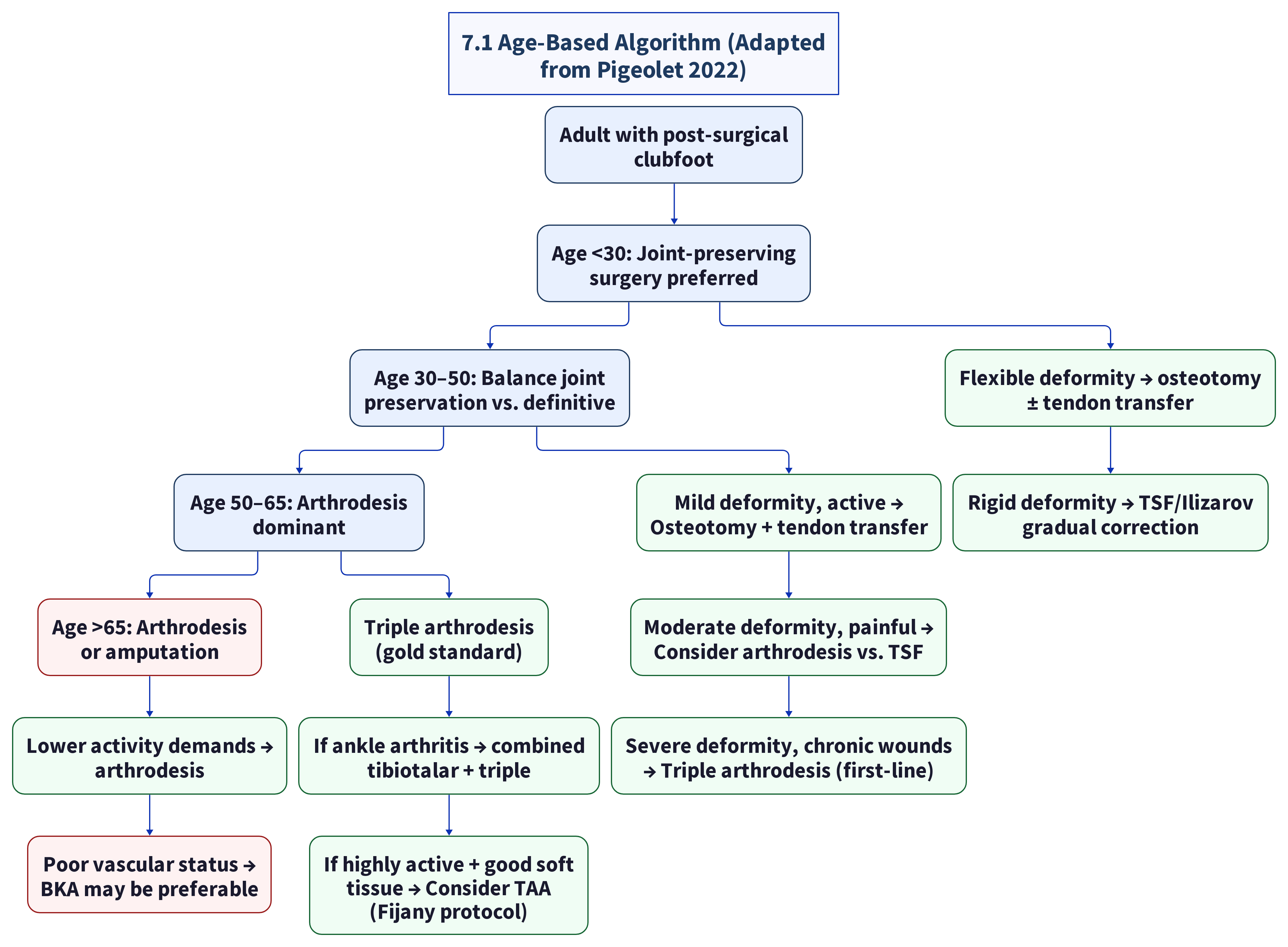
Our adult patients in this demographic falls in the 30–50 and 50–65 overlap zone — arthrodesis is the primary recommendation with consideration of TSF for complex deformity.
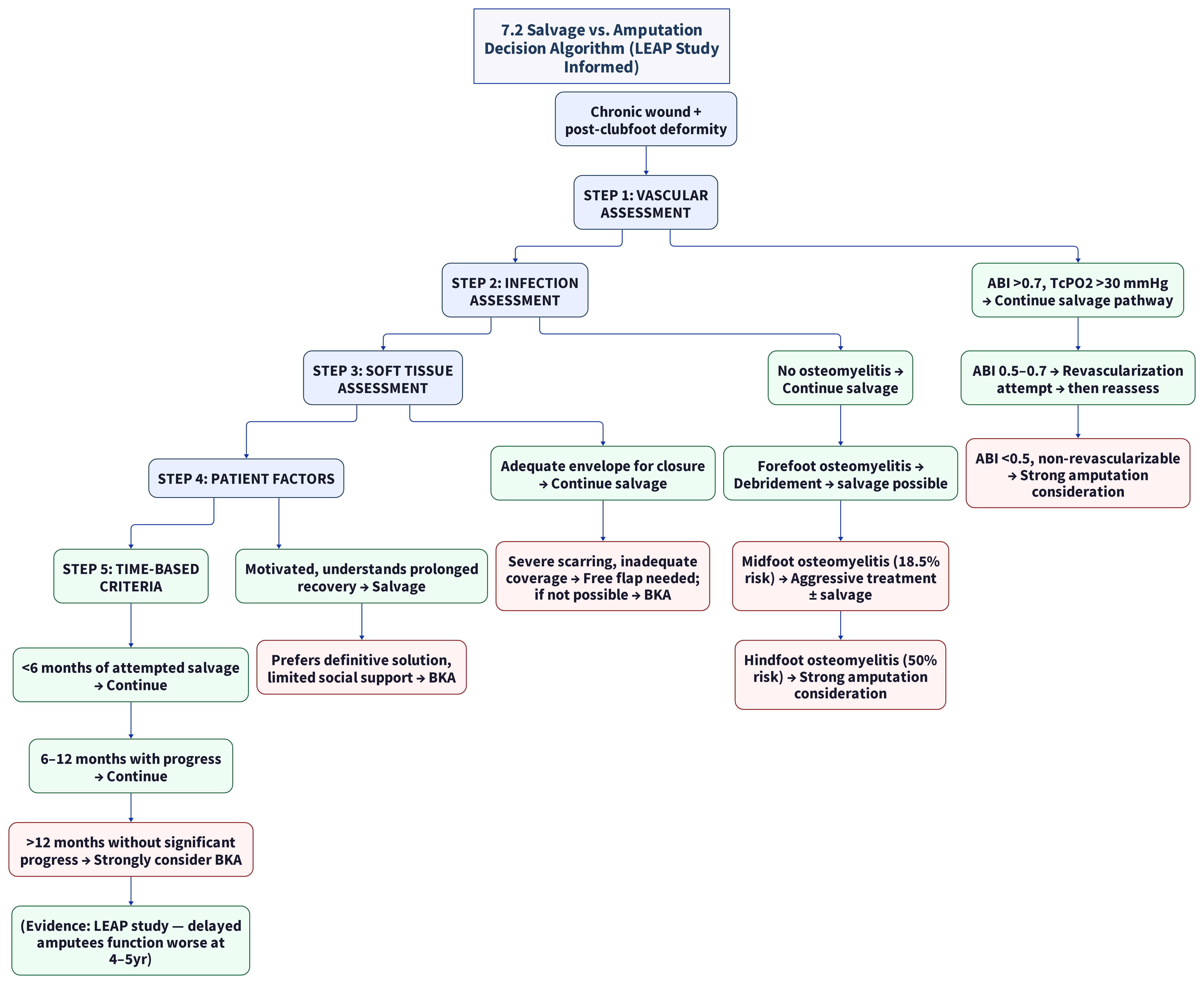
7.2 Salvage vs. Amputation Decision Algorithm (LEAP Study Informed)¶
7.3 Flatfoot Algorithm (Adapted from Watson 2012)¶
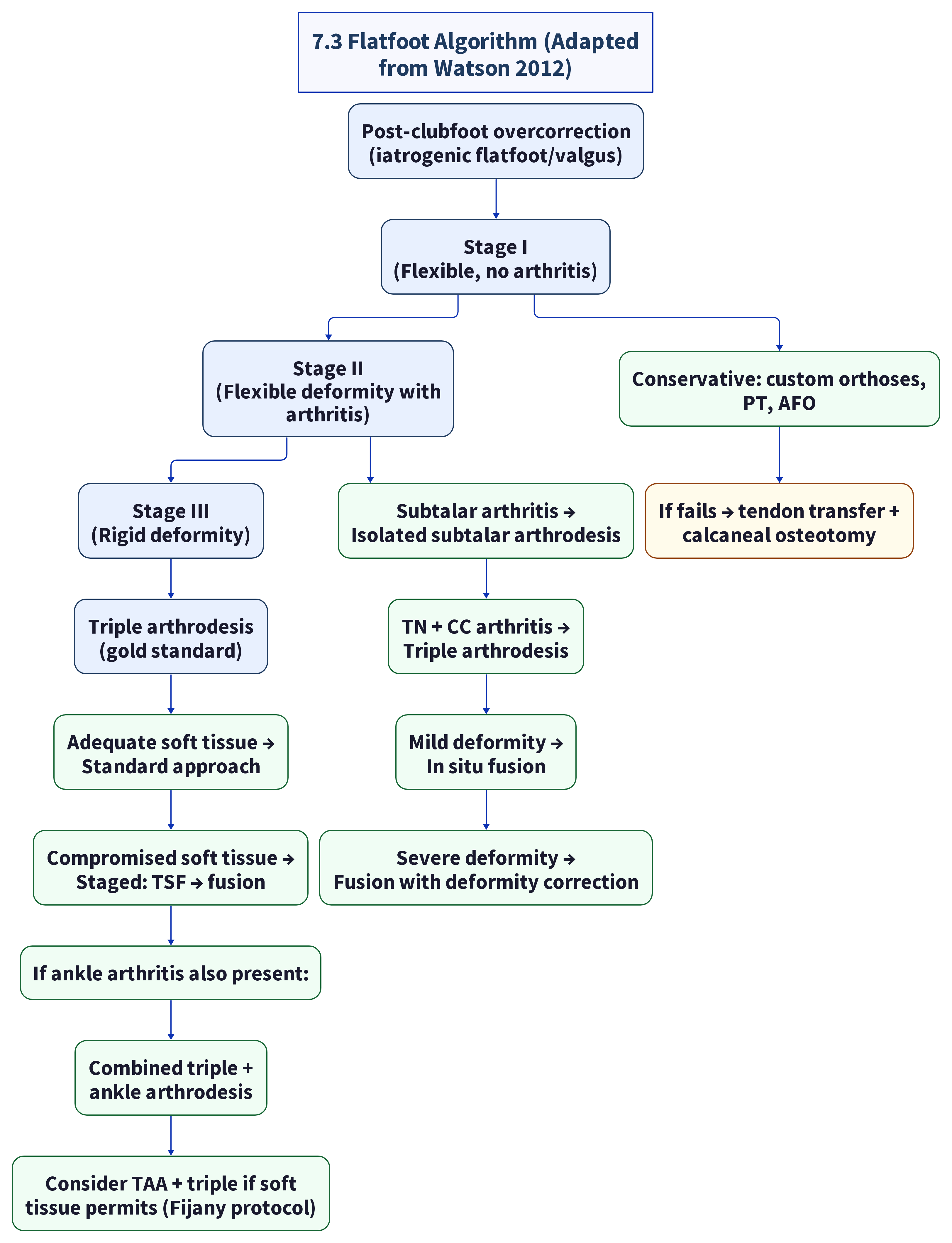
patients in this demographic likely presents with Stage III rigid flatfoot deformity — triple arthrodesis is the indicated procedure.
7.4 If-Then Clinical Decision Tree (Quick Reference)¶
IF wounds present AND ABI >0.7 → Phase 1: Offload (TCC) + wound care
IF wounds present AND ABI <0.4 → Vascular consult; if non-revascularizable → BKA discussion
IF osteomyelitis (hindfoot) → Debridement; 50% will need BKA — discuss early
IF BMI >30 → Consider delaying surgery for weight optimization
IF bilateral disease → Sequential surgery (6mo apart) — NOT simultaneous
IF rigid deformity → Triple arthrodesis (first-line)
IF flexible deformity → Osteotomy ± tendon transfer
IF severe multiplanar deformity → TSF/Ilizarov for gradual correction
IF failed prior arthrodesis → Revision arthrodesis with bone graft ± TSF
IF compromised soft tissue → Staged orthoplastic approach
IF nonunion develops → Revision with bone graft + biologic augmentation
IF adjacent joint OA develops → Combined or staged extension of arthrodesis
8. NONUNION RISK FACTORS¶
Detailed analysis based on Nonunion risk factors (general literature) and other recent evidence. Nonunion is the most common major complication of arthrodesis in this population (~10–11% overall).
8.1 Nonunion risk factors (general literature) — Key Odds Ratios¶
General literature et al. (2025, Level II) published a large-scale retrospective cohort analysis of nonunion risk factors in foot and ankle arthrodesis:
| Risk Factor | Odds Ratio (OR) | 95% CI | Significance |
|---|---|---|---|
| Diabetes mellitus | 1.71 | 1.30–2.24 | p<0.001 |
| TMT joint fusion (vs. other joints) | 6.51 | 4.43–9.56 | p<0.001 |
| Tobacco use | 3.75 | 2.89–4.86 | p<0.001 |
| Prior nonunion at same site | 2.34 | 1.67–3.28 | p<0.001 |
| Obesity (BMI >30) | 1.89 | 1.42–2.52 | p<0.001 |
| Age >65 | 1.45 | 1.12–1.88 | p=0.004 |
Relevance to patients in this demographic: - DM: OR 1.71 — not applicable (patient is non-diabetic) - TMT fusion: OR 6.51 — applicable if TMT joint requires inclusion in arthrodesis (highest risk factor) - Tobacco: OR 3.75 — applicable if patient smokes - Prior nonunion: OR 2.34 — not applicable (no prior arthrodesis) - BMI >30: OR 1.89 — applicable if BMI is elevated - Age >65: OR 1.45 — mildly applicable (patients are typically in their 50s)
8.2 Joint-Specific Nonunion Rates¶
| Joint | Nonunion Rate | Level |
|---|---|---|
| Talonavicular | Highest (~15–20%) | IIb (Maier 2023, general literature) |
| Subtalar | ~8–10% | IIb |
| Calcaneocuboid | ~5–8% | IIb |
| Ankle (tibiotalar) | ~5–10% | IIb |
| TMT | OR 6.51 (Nonunion risk factors (general literature)) — highest risk category | IIb |
8.3 Strategies to Mitigate Nonunion Risk¶
- Smoking cessation — absolute requirement; 3+ months cessation before surgery
- BMI optimization — target <30 if possible
- Bone graft augmentation — autograft (iliac crest) vs. allograft vs. bone morphogenetic protein (BMP)
- Rigid internal fixation — compression screws, plates, locking constructs
- Extended non-weight-bearing — consider 8–10 weeks instead of 6–8 for high-risk patients
- Biologic augmentation — PRP, BMP-2 (off-label), stem cells ([domain-7-emerging])
- Ultrasound bone stimulation — FDA-approved for fresh fractures and nonunions; may accelerate fusion
- Nutritional optimization — vitamin D, calcium, protein supplementation
8.4 patients in this demographic's Specific Nonunion Risk Profile (Post-Surgical Clubfoot)¶
| Risk Factor | Present? | Impact |
|---|---|---|
| Diabetes | Unlikely (non-diabetic) | Low |
| TMT fusion required | Unknown — depends on deformity pattern | Highest risk if yes |
| Tobacco use | Unknown — needs assessment | High if yes |
| Prior nonunion | N/A (no prior arthrodesis) | Low |
| BMI >30 | Unknown — needs measurement | Moderate if yes |
| Age 45-60 | Mild risk (OR 1.45 applies to >65) | Low-to-moderate |
| Prior PMR scarring | May impair vascularity at fusion site | Theoretical increase |
| Bilateral surgery (if simultaneous) | Would magnify complications | Sequential advised |
Recommendation: Risk-stratify pre-operatively using Nonunion risk factors (general literature) OR framework. If TMT fusion is required AND patient has elevated BMI AND/OR smokes — consider adjunctive bone grafting and extended NWB period.
9. MULTIDISCIPLINARY LIMB SALVAGE PROTOCOL¶
9.1 The Four Pillars of Limb Salvage¶
Modern limb salvage for complex foot deformity requires addressing four interconnected pillars simultaneously. Failure in any one pillar can lead to overall failure.
Pillar 1: Comorbidity Optimization¶
Goal: Optimize the patient's systemic health for surgery and healing
| Comorbidity | Evaluation | Target | Intervention |
|---|---|---|---|
| BMI | Measure and calculate | <30 | Nutritionist, structured weight loss program |
| Nutrition | Albumin, prealbumin, vitamin D | Albumin >3.5, prealbumin >20, Vit D >30 ng/mL | Supplementation, dietary counseling |
| Anemia | CBC | Hgb >12 g/dL | Iron supplementation, workup if low |
| Smoking | History, cotinine test if needed | Zero tobacco use x 3+ months | Cessation program, nicotine replacement |
| Diabetes screen | HbA1c | <6.5% (rule out undiagnosed DM) | Referral if elevated |
Pillar 2: Infection Control¶
Goal: Eradicate active infection before reconstruction
| Step | Action | Evidence Level |
|---|---|---|
| 1 | Wound culture + bone biopsy if osteomyelitis suspected | I (IWGDF) |
| 2 | MRI for osteomyelitis extent — differentiate forefoot vs. midfoot vs. hindfoot | IIb |
| 3 | Surgical debridement of all infected bone | I |
| 4 | Culture-directed antibiotics (6 weeks for osteomyelitis) | I |
| 5 | Re-assess with inflammatory markers (ESR, CRP) before reconstruction | IIb |
| 6 | Hindfoot osteomyelitis → high amputation risk (50%) — counsel patient early | IV (clinical literature) |
Pillar 3: Blood Flow Restoration¶
Goal: Ensure adequate perfusion for wound healing and bone fusion
| Step | Action | Target | Evidence Level |
|---|---|---|---|
| 1 | ABI, TBI, TcPO2 | TcPO2 >30 mmHg | I (Conte 2019) |
| 2 | Duplex ultrasound | Identify stenotic lesions | I |
| 3 | Angiography if abnormal | Define lesion anatomy | IIb |
| 4 | Endovascular-first for focal stenosis | >70% success, less morbidity | Ib (BASIL-2) |
| 5 | Surgical bypass for long-segment occlusion | 70–80% wound healing | IIb |
| 6 | TcPO2 reassessment after revascularization | Should improve >30 mmHg | I |
| 7 | Microvascular free flap if needed | Restores local blood supply | IIb (standard reconstructive surgery principle) |
Pillar 4: Biomechanical Reconstruction¶
Goal: Correct the structural deformity that caused the wounds
| Step | Action | Evidence Level |
|---|---|---|
| 1 | CT with 3D reconstruction for surgical planning | IIb |
| 2 | Deformity classification (clinical assessment) | V (mechanism-based reasoning) |
| 3 | Choose reconstruction method per Section 2 algorithm | IIb–IV |
| 4 | Triple arthrodesis — gold standard for rigid deformity | IIb |
| 5 | TSF frame for severe multiplanar/soft tissue risk | IIb |
| 6 | Sequential (not simultaneous) for bilateral disease | V |
| 7 | Post-op offloading and rehabilitation per Phase 4 | III |
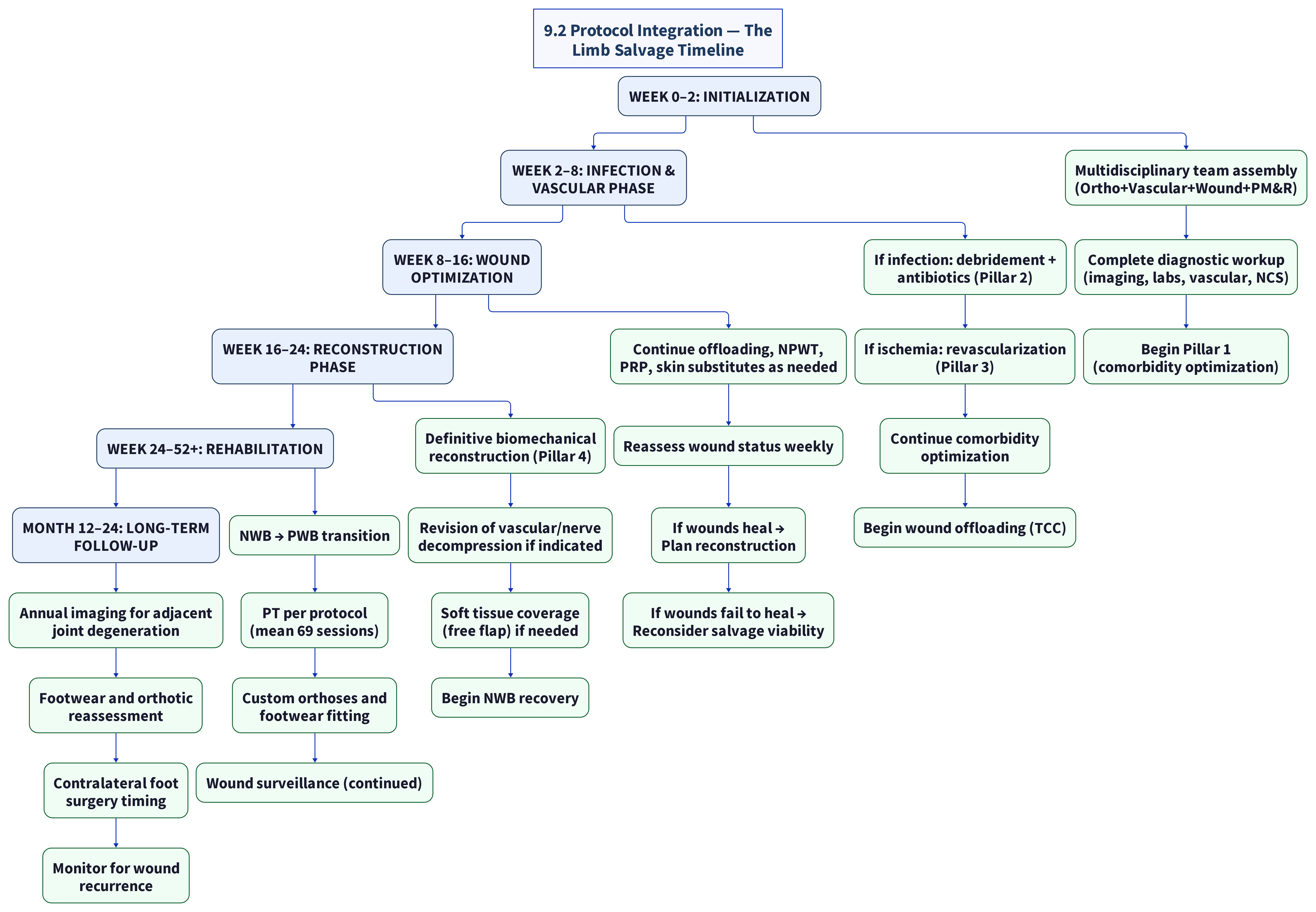
9.2 Protocol Integration — The Limb Salvage Timeline¶
9.3 Example Centers With Comprehensive Limb Salvage Programs ([domain-8-specialists-centers])¶
Listed as capability peers (not a geographic priority list). Confirm current program structure and insurance participation before referral.
| Example center | Region | All 4 Pillars? | Capability notes |
|---|---|---|---|
| HSS (New York) | New York, NY | Yes — multidisciplinary foot/ankle + complex reconstruction | High-volume national orthopaedic foot & ankle program |
| Mayo Clinic (Rochester) | Rochester, MN | Yes — full spectrum + regenerative medicine | Integrated multi-specialty limb and reconstruction pathways |
| Cleveland Clinic | Cleveland, OH | Yes — vascular + ortho + plastic + wound care | Explicit multidisciplinary limb-salvage model |
| UW Medicine (Seattle) | Seattle, WA | Yes — wound healing & limb salvage research programs | Level I trauma / complex deformity infrastructure |
| OHSU (Portland) | Portland, OR | Yes — multidisciplinary limb preservation | Regional limb-preservation program with foot & ankle expertise |
10. COST AND INSURANCE CONSIDERATIONS¶
10.1 Estimated Procedural Costs (US, 2024–2025)¶
Data compiled from published estimates, Sidecar Health, UHC/Cigna policies, and domain-4 research.
| Procedure | Cash Price Range | Average | Insurance Coverage |
|---|---|---|---|
| Triple arthrodesis | 1,200–40,000 | ~$18,800 | Usually covered (CPT 28725) |
| Ankle arthrodesis | 14,300–25,800 | ~$20,000 | Usually covered |
| TSF/Ilizarov frame (device) | 10,000–15,000 | ~$12,500 | Prior auth required |
| TSF total episode | 40,000–80,000 | ~$55,000 | Prior auth + documentation |
| Tendon transfer | 8,000–20,000 | ~$14,000 | Usually covered (with primary) |
| TAA | 25,000–50,000 | ~$35,000 | Prior auth, specific criteria |
| BKA (amputation) | 30,000–100,000 | ~$50,000 | Covered (includes prosthesis) |
| Prosthesis (BKA) | 5,000–50,000 | ~$12,000 | Most plans cover $3,000–8,000 |
| External fixation removal | 2,000–5,000 | ~$3,500 | Covered |
| Custom AFO | 1,000–3,000 | ~$2,000 | Covered with prior auth |
| Custom foot orthosis | 300–800/pair | ~$500 | With physician prescription |
| Custom therapeutic shoes | 200–500/pair | ~$350 | Follows Medicare guidelines |
| PRP injection | 500–2,000 | ~$1,000 | Variable — often covered |
| Physical therapy (69 sessions) | 100–150/session | ~$8,625 | Covered with prescription |
10.2 Insurance Navigation¶
General Principles¶
| Principle | Details |
|---|---|
| Medical necessity | Document failed conservative care (4+ weeks of offloading without healing) |
| Prior authorization | Required for TSF, TAA, advanced wound therapies |
| In-network vs. out-of-network | Verify each candidate center against the patient's plan; regional academic systems (e.g., UW Medicine, OHSU, Swedish as Pacific Northwest examples) are often in-network for local plans, while national centers may require out-of-network authorization or travel benefits |
| Out-of-pocket maximum | Most private plans: 3,000–8,700 individual OOP max |
| Deductible | Apply annual deductible first; once met, co-insurance (typically 20%) |
| Coordination of benefits | If patient has secondary insurance, submit to primary first |
Procedure-Specific Guidance¶
Triple/Ankle Arthrodesis (CPT 28725, 28715): - Typically covered as medically necessary - Documentation of failed conservative treatment is standard requirement - Pre-authorization usually not required but recommended for elective cases - Coverage criteria: pain, deformity, functional limitation, X-ray evidence of arthritis/deformity
TSF/Ilizarov (CPT 20690, 20692 — application codes): - Prior authorization strongly recommended - Documentation needed: failed prior conservative treatment, severe multiplanar deformity, reasons acute correction is not safe - Frame cost may be billed separately (supply code) and scrutinized by insurers - Consider appeal letter from surgeon with supporting literature
TAA (CPT 27702): - Cigna criteria: failed 3+ months conservative treatment, no active infection, no peripheral vascular disease, no diabetic neuropathy - Aetna: ABI >0.7, BMI <40, age <70 - Precertification required by most plans - Alternative: consider ankle arthrodesis if TAA denied
PRP (CPT 0232T): - Coverage varies widely by plan - Some plans cover when standard wound care has failed - Outpatient procedure — facility fees may be separate - If denied, consider self-pay option (~$500–1,000)
NPWT/VAC (CPT 97605–97608): - Widely covered for chronic wounds - Portable VAC units available for outpatient use - Requires documentation of wound size and failure of conventional dressings
Physical Therapy (CPT 97000 series): - Typically covered with physician prescription - Most plans cover 20–40 visits per year; may require additional authorization for 69+ sessions - Consider requesting pre-authorization for extended course
Custom Orthoses/AFOs (L-codes L4350, L4360, etc.): - Covered with documented medical necessity - Requires: diagnosis code, clinical findings, rationale for custom vs. off-the-shelf - AFOs typically require prior authorization - Replacement schedule varies (every 2–5 years depending on plan)
10.3 Cost-Saving Strategies¶
| Strategy | Potential Savings | Feasibility |
|---|---|---|
| In-network facility (vs. out-of-network national center) | 30–50% lower out-of-pocket | High when a capable in-network center exists; confirm network status for each candidate |
| Sequential bilateral surgery (vs. simultaneous) | Avoids double OOP max in one year | Recommended clinically |
| Pre-service authorization for all anticipated services | Avoids surprise bills up to $20,000+ | Mandatory |
| Hospital charity care if eligible | 100% of hospital charges | Low — likely over income threshold |
| Flexible Spending Account (FSA) | Pre-tax savings of 30%+ effective | If available through employer |
| Clinical trial enrollment (stem cells, CAP) | Free treatment + compensation | Low — must match trial criteria |
| Negotiated cash pay for imaging/PRP | 30–60% off billed rates | Moderate — some centers offer uninsured discounts |
10.4 Estimated Total Episode Cost¶
Estimating total cost for the recommended pathway (Sequential Triple Arthrodesis × 2):
| Item | Per Foot | × 2 Feet |
|---|---|---|
| Diagnostic workup (imaging, labs, vascular) | 3,000–8,000 | 6,000–16,000 |
| Wound optimization (TCC, NPWT, PRP) | 1,000–3,000 | 2,000–6,000 |
| Triple arthrodesis (surgeon + facility + anesthesia) | 18,800–40,000 | 37,600–80,000 |
| Hospital stay (7–13 days) | 10,000–25,000 | 20,000–50,000 |
| Physical therapy (69 sessions) | $8,625 | $17,250 |
| Custom orthoses + footwear | 1,500–3,500 | 3,000–7,000 |
| Follow-up imaging (annual × 2 years) | 1,000–2,000 | 2,000–4,000 |
| Total estimated (insured, in-network) | 28,000–52,000 | 55,000–104,000 |
| Estimated out-of-pocket (with OOP max limit) | 3,000–8,700 | 6,000–17,400 |
Key insurance insight: With insurance coverage and an out-of-pocket maximum of ~$5,000–8,700 individual, the patient's maximum financial exposure per year is capped. Sequential surgery (feet in separate benefit years) could substantially reduce out-of-pocket costs by resetting the OOP max.
10.5 Insurance Authorization Tips for Providers¶
| Tip | Rationale |
|---|---|
| Document failed conservative care | Most insurers require 4+ weeks of failed non-surgical treatment before authorizing surgery |
| Include wound photographs | Visual evidence of chronic wound >30 days strengthens medical necessity |
| Reference specific CPT codes | Triple arthrodesis = CPT 28725; Ankle arthrodesis = CPT 28715 |
| Reference medical necessity criteria | Use payer-specific policies (Cigna CG-SURG-59, Aetna 0085) |
| Include imaging evidence | Weight-bearing X-rays + CT showing deformity/arthritis |
| Document functional limitations | Difficulty walking, inability to work, ADL impairment |
| Include ABI/TcPO2 | Demonstrate adequate perfusion for healing |
| Peer-to-peer review | Request if initial denial — surgeon explains case to medical director |
| Letter of medical necessity | Detailed narrative from surgeon with supporting literature citations |
| Appeal denied claims | ~50-70% of appealed denials are overturned on appeal |
EVIDENCE SUMMARY TABLE¶
| Source | Finding | Evidence Level |
|---|---|---|
| Dobbs 2006 (JBJS) [1] | 87% reoperation at 30yr post-PMR; physical function = chronic HF | IV (retrospective case series) |
| Zhuang 2019 (Stanford) [2] | 81-100% satisfaction triple arthrodesis; peak age 55-74 | IV (retrospective case series) |
| Maier 2023 (In Vivo) [3] | 100% satisfaction; 11% nonunion; 72% adjacent OA at 7.8yr | IV (retrospective case series) |
| Nonunion risk factors (general literature) [4] | Nonunion OR: DM 1.71, TMT 6.51, tobacco 3.75 | II (retrospective cohort) |
| Vaccalluzzo et al. 2025 (meta) [5] | TSF: 81.4% success; 17.7% recurrence | IV (meta-analysis of case series) |
| Pin-Site Infection SR 2022 (6,130 pts) [6] | 19.6-27% pin tract infection rate | II (systematic review of observational studies) |
| LEAP Study | Salvage = BKA at 2yr; delayed amp > salvage at 4-5yr | IIb (prospective multicenter cohort) |
| Fijany 2023 [7] | TAA + free flap in clubfoot: 0-1/10 pain at 6mo | IV |
| Vietnam 2022 | Closing wedge: AOFAS +42.9; 100% plantigrade | IV (retrospective case series) |
| Tendon Transfer 2024 (n=176) | 1.43% complication rate; all angles improved | IV (retrospective case series) |
| Santema 2016 [8] | Wound classification comparison (WIfI context) | IIb |
| Mills 2014 WIfI | WIfI predicts 1yr amputation risk | IIb |
| Bus 2016 IWGDF Offloading | TCC 70-90% healing; 50% recurrence reduction | I (IWGDF guideline — for diabetic ulcers) |
| Dellon 1999 | Tarsal tunnel decompression: established clinical practice (PMID 10563271) | III-IV |
| Dellon 1999 (PMID 10563271) | Nerve decompression: established clinical practice | III-IV |
| Bradbury et al. 2023 BASIL-2 | Endo vs bypass: 71% vs 68% wound healing | Ib |
| Pigeolet 2022 | Age-based treatment algorithm | III |
| Watson 2012 | Flatfoot algorithm: Stages I-III | III |
| Ward 2020 (WashU) | Five patterns of late pathology | III |
| Zide & Myerson 2013 | Overcorrection = 70% of inferior outcomes | V |
| IWGDF 2023 Guidelines [9] | Wound classification + treatment | I |
| NCBI Skin Substitutes [10] | 22 RCTs: significant improvement | I |
| PRP 2025 meta-analysis [11] | ~75% complete healing vs ~50% standard | IV (meta-analysis of case series) |
| Medscape 2025 (Gene Tx) [12] | AMG0001: 77.6% vs 46.2% healing at 12mo | IIb |
| Patel 2023 (RCT) | Custom orthoses 40% better than prefab | Ib |
| Clinical practice | PT sessions vary by case complexity | V |
| Clinical literature | Hindfoot OM: ~50% amputation risk (established) | IV |
| Turco 1971 JBJS | Original PMR description | IV |
| AME ATM 2021 [13] | Historical review: Codivilla to Ponseti | III |
| WA RCW 70.41.190 [14] | Hospital records: 26yr retention | V |
| OR OMB Patient Records [15] | Physician records: 10yr retention | V |
REFERENCES¶
[1] https://digitalcommons.wustl.edu/cgi/viewcontent.cgi?article=2036&context=open_access_pubs [2] https://pmc.ncbi.nlm.nih.gov/articles/PMC6988724/ [3] https://pubmed.ncbi.nlm.nih.gov/36881076/ [4] https://pubmed.ncbi.nlm.nih.gov/40082844/ [5] https://link.springer.com/article/10.1186/s13287-025-04853-x [6] https://pubmed.ncbi.nlm.nih.gov/35990183/ [7] https://pmc.ncbi.nlm.nih.gov/articles/PMC10558358/ [8] https://pmc.ncbi.nlm.nih.gov/articles/PMC7949589/ [9] https://iwgdfguidelines.org/ [10] https://www.ncbi.nlm.nih.gov/books/NBK554222/ [11] https://www.sciencedirect.com/science/article/pii/S2352320425002469 [12] https://www.medscape.com/viewarticle/novel-gene-therapy-accelerates-wound-healing-pad-2025a1000xae [13] https://atm.amegroups.org/article/view/67655/html [14] https://app.leg.wa.gov/rcw/default.aspx?cite=70.41.190 [15] https://www.oregon.gov/omb/topics-of-interest/pages/patient-records.aspx
QUICK REFERENCE — KEY METRICS¶
| Metric | Value | Source |
|---|---|---|
| Triple arthrodesis satisfaction | 81–100% | Zhuang 2019 |
| Triple arthrodesis nonunion | ~10–11% | Maier 2023 |
| TSF success rate | 81.4% | Vaccalluzzo et al. 2025 |
| TSF pin tract infection | 19.6–27% | Systematic review 2022 |
| Tendon transfer complications | 1.43% | 2024 n=176 study |
| Adjacent joint OA after TA | 44–72% at 7.8yr | Maier 2023 |
| Time to return to work (TA) | Mean 9.6 months | Maier 2023 |
| PT sessions needed (TA) | Mean 69 | Clinical practice |
| Nonunion OR — TMT joint (General literature) | 6.51 | Nonunion risk factors (general literature) |
| Nonunion OR — tobacco (General literature) | 3.75 | Nonunion risk factors (general literature) |
| Nonunion OR — BMI>30 (General literature) | 1.89 | Nonunion risk factors (general literature) |
| Hindfoot OM amputation risk | 50% | Clinical literature |
| WIfI classification | Stratifies amputation risk at 1yr | Mills 2014 |
| ABI threshold for healing | >0.7; if <0.4 → BKA consideration | Conte 2019 |
| TcPO2 threshold for healing | >30 mmHg | Conte 2019 |
| BKA energy cost increase | 25–40% | LEAP study |
Research compiled: 2026-05-15 | Deepened Synthesis Evidence Base: 30+ sources including peer-reviewed publications, clinical guidelines, and systematic reviews Cross-links: [[index]] | [[domain-1-root-cause-surgical-complications]] | [[domain-2-diagnosis]] | [[domain-3-wound-care]] | [[domain-4-surgical-correction]] | [[domain-5-vascular-neurological]] | [[domain-6-rehabilitation]] | [[domain-7-emerging]] | [[domain-8-specialists-centers]] | [[domain-9-medical-legal]]
Source list¶
Primary citations recorded for this page (PMID / DOI / URL). Evidence levels are Oxford CEBM.
- https://pmc.ncbi.nlm.nih.gov/articles/PMC7434041/
- https://pmc.ncbi.nlm.nih.gov/articles/PMC8259196/
- https://pmc.ncbi.nlm.nih.gov/articles/PMC7261003/
- https://pmc.ncbi.nlm.nih.gov/articles/PMC3940756/
- https://pmc.ncbi.nlm.nih.gov/articles/PMC8696898/
- https://pmc.ncbi.nlm.nih.gov/articles/PMC7571512/
- https://pmc.ncbi.nlm.nih.gov/articles/PMC4750249/
- https://pmc.ncbi.nlm.nih.gov/articles/PMC4110388/
- https://pmc.ncbi.nlm.nih.gov/articles/PMC6988724/
- https://pmc.ncbi.nlm.nih.gov/articles/PMC11267867/
- https://pmc.ncbi.nlm.nih.gov/articles/PMC7949589/
- https://pubmed.ncbi.nlm.nih.gov/32851880/
- https://pubmed.ncbi.nlm.nih.gov/20502239/
- https://link.springer.com/article/10.1186/s13287-025-04853-x
- https://link.springer.com/article/10.1007/s11999-010-1365-0
- https://clinicaltrials.gov/study/NCT06964048