Domain 5 — Vascular & Neurological Complications in Adult Post-Clubfoot
Status: Research Synthesis Clinical Focus: Adult post-surgical clubfoot complications — chronic wounds, progressive deformity, and mobility loss following childhood corrective surgery General Clinical Reference Last Updated: 2026-05-16
This domain examines how vascular insufficiency and peripheral neuropathy from prior clubfoot surgeries contribute to wound non-healing, and what vascular and neurological interventions can improve outcomes. This is directly relevant to the patient's chronic wounds that fail to heal despite standard wound care [[domain-3-wound-care]].
Cross-links: [[index]] | [[domain-1-root-cause-surgical-complications]] | [[domain-3-wound-care]] | [[domain-4-surgical-correction]] | [[domain-7-emerging]] | [[domain-8-specialists-centers]]
5.1 Pathophysiological Framework: The Vascular-Neurological-Surgical Intersection
How Prior PMR Surgery Compromises Vascular Supply
Childhood posteromedial release (PMR) procedures in the 1970s-1990s created long-term vascular consequences that manifest decades later as poor wound healing:
- Direct vascular injury during surgery: The posterior tibial artery runs directly through the posteromedial diszone. Extensive PMR procedures risked: (unverified)
- Partial transection or cautery injury to the posterior tibial artery and its branches
- Saphenous vein harvest or injury compromising venous drainage
-
Intimal damage from surgical retraction creating delayed stenosis
-
Scar tissue and fibrosis: Extensive soft-tissue dissection creates: (unverified)
- Dense scar tissue that entraps and compresses small vessels
- Fibrosis that reduces capillary density and microvascular perfusion
-
Loss of perivascular tissue support leading to vascular fragility
-
Altered anatomical relationships: Overcorrection and subsequent deformity changes: (unverified)
- Vascular routing and tension on arterial segments
- Venous return pathways (calcaneal position affects venous pump efficiency)
- Lymphatic drainage contributing to chronic edema which further impairs perfusion
How Neuropathy Compounds Wound Non-Healing
| Mechanism | Pathophysiology | Clinical Consequence |
|---|---|---|
| Large fiber neuropathy | Compression/stretch of tibial nerve from hindfoot valgus/varus | Loss of protective sensation → unrecognized trauma |
| Small fiber neuropathy | Damage to C-fibers and Aδ-fibers from scar tissue | Neuropathic pain → altered weight-bearing → pressure points |
| Autonomic neuropathy | Sympathetic fiber disruption from surgery or compression loss of sudomotor, vasomotor, pilomotor function | Dry skin → fissuring → portal for infection; impaired vasodilation during healing |
| Tarsal tunnel syndrome | Tibial nerve compression at flexor retinaculum from deformity | Burning, tingling, plantar numbness; may be bilateral |
| Peroneal nerve entrapment | Compression at fibular neck from altered gait and compensatory mechanics | Foot drop, decreased dorsiflexion power |
(unverified)
The Vicious Cycle of Non-Healing
PMR Surgery (1970s-90s)
 ↓
Vascular compromise (macro + micro) + Nerve injury/entrapment
 ↓
Reduced perfusion + Loss of protective sensation
 ↓
Micro-trauma from abnormal loading + impaired skin integrity
 ↓
Ulceration with poor healing response
 ↓
Chronic wound → infection → osteomyelitis risk
 ↓
Further tissue destruction → escalating wound severity
5.2 Vascular Assessment in the Post-Clubfoot Foot
Clinical Assessment Protocol
| Test | Purpose | Normal Value | Abnormal Threshold |
|---|---|---|---|
| Ankle-Brachial Index (ABI) | Macrovascular assessment | 1.0–1.3 | <0.9 = PAD; >1.3 = calcified/uncompressible |
| Toe-Brachial Index (TBI) | Microvascular assessment (unaffected by calcification) | >0.7 | <0.7 = impaired digital perfusion |
| Transcutaneous Oxygen (TcPO2) | Tissue-level oxygenation | >40 mmHg | <30 mmHg = unlikely to heal; <20 mmHg = critical ischemia |
| Stimulation TcPO2 | Dynamic assessment of local microvascular reserve | Normal >35 mmHg under stimulation | <25 mmHg under stimulation = impaired healing; may identify PAD missed by resting TcPO2 [[PMC8704582]] |
| Duplex Ultrasound | Arterial stenosis mapping | Peak systolic velocity ratio <2.0 | Ratio >2.0 = >50% stenosis; >4.0 = >80% stenosis |
| CT/MR Angiography | Detailed vascular mapping | Patent vessels | Stenosis, occlusion, collateral status |
| Skin Perfusion Pressure (SPP) | Microvascular perfusion | >50 mmHg | <30 mmHg = impaired healing likely |
| Nerve Conduction Studies (NCS) | Large fiber neuropathy quantification | Normal conduction velocity | Reduced amplitude/velocity = neuropathy |
WIfI Classification System (Society for Vascular Surgery) (verified against PubMed) PMID 24126108
The Wound, Ischemia, and foot Infection (WIfI) classification system stratifies limbs by amputation risk and revascularization benefit:
| Grade | Wound | Ischemia | Infection |
|---|---|---|---|
| 0 | No ulcer | None (ABI ≥0.9, TBI ≥0.7) | None |
| 1 | Small, shallow ulcer | Mild (ABI 0.6-0.9, TBI 0.4-0.6) | Mild |
| 2 | Moderate tissue loss | Moderate (ABI 0.4-0.6, TBI 0.2-0.4) | Moderate |
| 3 | Extensive/gangrene | Severe (ABI <0.4, TBI <0.2) | Severe |
Clinical relevance: For adult patients with chronic wounds and compromised vascular supply from prior PMR, a WIfI assessment provides objective data for insurance authorization of vascular interventions and surgical reconstruction planning [[domain-4-surgical-correction]].
Evidence Summary for Vascular Assessment
| Study | Finding | Level |
|---|---|---|
| Mills et al. 2014 (WIfI Validation) (verified against PubMed) PMID 24126108 | WIfI predicts amputation risk and revascularization benefit | IIb |
| Conte et al. 2019 (Global Vascular Guidelines) (verified against PubMed) | Comprehensive algorithm for CLTI evaluation and management | I |
| Cerqueira et al. 2020 (WIfI Review, PMC8202158) (PMID 34178056) | WIfI classification literature review: system predicts amputation risk and guides revascularization decisions | IIb |
| Beropoulis et al. 2016 (PMID 26994958) | WIfI classification predicted amputation risk in nondiabetic CLTI patients treated by endovascular therapy; demonstrated applicability beyond diabetic populations | IIb |
| Robinson et al. 2024 (J Vasc Surg) (PMID 28410924) | Review of WIfI clinical staging confirms predictive ability across diverse populations; recommended as standard language for limb threat assessment | III |
| Darcey et al. 2022 (EJVES) | WIfI-based risk factor analysis in CLTI patients confirms independent predictive value of ischemia grading component | IIb |
| Sumpio 2020 | Non-diabetic vascular insufficiency causes 12-15% of chronic foot ulcers | III |
| Rajbhandari et al. 2020 (PMID 35208490) | Neuropathic foot ulcers occur in non-diabetic patients, often post-surgical | III |
5.3 Vascular Interventions for Wound Healing
Endovascular Therapy (Angioplasty/Stenting)
Evidence: Endovascular intervention has become first-line for many cases of peripheral arterial disease affecting wound healing.
| Study | Technique | N | Wound Healing Rate | Level |
|---|---|---|---|---|
| Bradbury et al. 2023 (BASIL-2 trial, Lancet) (PMID 37116524) | Endovascular-first vs. vein bypass-first | 219 (IP cohort) | Endo-first was SUPERIOR to bypass-first for IP revascularization (35% reduced major amputation/death risk) | Ib |
| Bhamidipaty et al. 2019 (PMID 34251741) | Endovascular revascularization for CLTI limb salvage meta-analysis | 28 studies | 78% wound healing at 12 months | IIa |
| Conte et al. 2019 (verified against PubMed) | Global guidelines recommend endovascular where anatomy favorable | Expert consensus | I | |
| Menard et al. 2024 (PMID 39069016, PMC11734614, BEST-CLI Trial) (verified against source) | Revascularization strategy significantly impacts CLTI outcomes; open bypass superior to endovascular for clinical failure, hemodynamic failure, and symptom resolution (P<.001) | Ib |
Clinical application for adult post-surgical clubfoot patients: - If duplex/angiography reveals discrete stenosis of posterior tibial or dorsalis pedis arteries (from prior surgical scarring or intimal injury) - Balloon angioplasty ± stent placement may improve perfusion to the wound bed - TcPO2 improvement from <30 to >30 mmHg is a strong predictor of wound healing (verified against PubMed) - Insurance consideration: Insurance typically covers endovascular procedures for documented PAD with chronic wounds
Surgical Bypass
| Study | Finding | Level |
|---|---|---|
| BASIL-2 (Bradbury et al. 2023) | Bypass-first strategy was associated with 35% increased risk of major amputation or death compared to endo-first for infrapopliteal revascularization | Ib |
| Norgren et al. 2007 (TASC II) (verified against PubMed) PMID 17489079 | Bypass preferred for long-segment occlusions (TASC C/D lesions) | IIb |
| Darling et al. 2025 (PMID 39826656, BEST-CLI Validation) | Vein graft bypass achieved 47% complete wound healing at 6 months vs 40% endovascular; 53% reduction in major reintervention at 7 years | Ib |
| Chamseddine et al. 2025 (PMID 40769463) | Popliteal-distal bypass restores direct flow to tibial/foot arteries for CLTI secondary to infrapopliteal occlusive disease | III |
Considerations for post-clubfoot foot: - Prior surgical scarring may complicate distal anastomosis site selection (verified against PubMed) - Vein graft availability must be assessed (prior leg surgeries may have compromised saphenous vein) (verified against PubMed) - Bypass to dorsalis pedis or plantar arteries technically demanding but feasible in experienced centers (verified against PubMed) - Wound healing rates of 70-80% achieved with successful bypass to patent pedal vessels (verified against PubMed) PMID 26870158
Lumbar Sympathectomy
Mechanism: Sympathetic nerve interruption causes vasodilation of skin microcirculation, increasing blood flow to ischemic skin and subcutaneous tissue. (verified against PubMed)
| Study | Technique | N | Wound Healing Rate | Level |
|---|---|---|---|---|
| Nehler et al. 2004 (unverified) | Catheter-based lumbar sympathectomy for non-reconstructable PAD | 54 legs | 45% complete wound healing, 62% improvement in rest pain | III |
| Meta-analysis 2018 (J Vasc Surg) (unverified) | Chemical vs. surgical sympathectomy | 791 patients | 39% complete healing (chemical) vs 44% (surgical) | IIb |
| Bipolar RF LSB (2025, Wound Repair Regen) (unverified) | Bipolar radiofrequency lumbar sympathetic block for CLTI | 60 patients | 55% complete wound healing vs 30% in control group (p < 0.05); statistically significant improvement | IIb |
| CT-Guided Lumbar Sympathectomy (AJR 2021) (unverified) | CT-guided percutaneous chemical (alcohol/phenol) sympathectomy | 87 patients | 30-87% success rates; complication risk <1% | IIb |
| Shaalan et al. 2024 (PMC11542267) (PMID 39511575) | Retroperitoneal laparoscopic sympathectomy | 24 patients (not 142) | Laparoscopic: shorter stay, fewer complications; equivalent outcomes | IIb |
| Wardak et al. 2026 (PMID 41254237) | Systematic review of 16 studies: lumbar sympathectomy provides pain relief, wound healing, and quality-of-life improvement when revascularization is not feasible | IV |
Updated evidence (2024-2025): Recent advances in lumbar sympathectomy include: - Bipolar radiofrequency ablation (Wound Repair Regen 2025): (unverified) - CT-guided chemical sympathectomy (AJR 2021): (unverified) - Laparoscopic sympathectomy (Shaalan et al. 2024, PMC11542267): (verified against PubMed) PMID 39511575
Indications for adult post-surgical clubfoot patients: - Last resort when revascularization not possible (no target vessels, diffuse small vessel disease) - Most effective for rest pain and skin perfusion; limited benefit if macrovascular occlusion present - CT-guided chemical sympathectomy (alcohol or phenol) is the least invasive option - Bipolar RF may offer best current wound healing rates for appropriate candidates (unverified) - Can be combined with medical therapy (cilostazol, pentoxifylline) for additive effect (verified against PubMed)
Microvascular Reconstruction
| Study | Technique | N | Outcome | Level |
|---|---|---|---|---|
| Shimbo et al. 2023 (PMID 36577499) | Meta-analysis of 15 studies: combined revascularization and free flap for CLTI — 91% flap survival, 86% 1-year limb salvage, 81% 3-year limb salvage | IIa | ||
| Wortman et al. 2023 (PMID 37809200) | Latissimus dorsi free flap for orthoplastic reconstruction in relapsed clubfoot enabling TAA | 1 | Successful TAA in compromised soft-tissue envelope | IV |
| Fitzgerald O'Connor et al. 2011 (PMID 21163675) | Systematic review of 528 patients: free tissue transfer for non-traumatic lower extremity wounds — 92% flap survival, 83.4% limb salvage | IIa | ||
| Arakelyan et al. 2022 (PMC8779818) (PMID 35111561) | Retrospective evaluation of 182 free flaps in extremity reconstruction | 182 flaps | Flap survival rate ~83% (total flap failure 17.03%); flaps performed day 4-14 had lowest failure rate (p=0.022) | III |
Application for adult post-surgical clubfoot patients: If chronic wounds result from structural deformity with inadequate soft tissue coverage, microvascular free flap reconstruction may be needed after vascular optimization and before definitive joint reconstruction [[domain-4-surgical-correction]].
Timing considerations: Free flap reconstruction in the post-clubfoot foot should be planned as part of a staged approach: 1. Phase 1: Vascular optimization (endovascular or bypass) to improve inflow to the recipient site 2. Phase 2: Wound debridement + simultaneous free flap transfer for coverage 3. Phase 3: Definitive bony reconstruction (arthrodesis or osteotomy) once soft-tissue envelope is healed and infection-free 4. Phase 4: Delayed secondary procedures (tendon transfers, nerve decompression) as needed
(unverified)
Tarsal Tunnel Syndrome in Post-Clubfoot Adults
Pathophysiology: The tarsal tunnel is formed by the flexor retinaculum extending from medial malleolus to calcaneus. In post-clubfoot feet, several factors increase compression risk:
- Hindfoot valgus (from overcorrection) stretches the tibial nerve across the tunnel
- Hindfoot varus (from undercorrection) compresses the nerve against the medial wall
- Post-surgical scar tissue from PMR creates adhesions around the nerve
- Ganglion cysts or hypertrophied muscles secondary to altered mechanics
- Chronic inflammation from abnormal mechanics causes tunnel fibrosis
(unverified)Clinical presentation: - Burning/tingling in plantar foot (medial and lateral plantar nerve distribution) - Night symptoms, worse with prolonged standing - Positive Tinel's sign at medial malleolus - Reduced sensation in plantar distribution - Pain radiating to medial heel or toes
(verified against PubMed)
Evidence for Nerve Decompression
| Study | Technique | N | Success Rate | Level |
|---|---|---|---|---|
| Dellon 1999/2015 (unverified) | Surgical decompression of tarsal tunnel + multiple lower extremity nerve compression sites | 300+ | 80-85% significant symptom relief | III |
| Boschetti et al. 2013 (unverified) | Systematic review of tarsal tunnel decompression outcomes | 12 studies, 438 patients | 74-94% improvement (pooled ~85%) | IIb |
| Haq et al. 2024 (PMC11296061) (PMID 39101044) | Scoping review of tarsal tunnel management | 32 studies (not 40 as claimed) | 75.3% excellent/good results overall; 24.7% fair/poor (verified against PubMed) | IIb |
| Casares Tamayo et al. 2026 (PMC13090096) (PMID 42003951) | Surgical decompression for TTS — functional outcomes assessment | 15 patients (not 68) | AOFAS and VAS outcomes reported; specific "70-90% success" claim NOT verified from abstract | III |
| Endoscopic TTS Decompression (2013) (unverified) | Endoscopic vs open tarsal tunnel decompression systematic review | Pooled data | Endoscopic: lower complication rate, faster return to ambulation; success rates 44-96% depending on patient selection | IIb |
| Schaper et al. 2019 (unverified) | Nerve decompression in patients with prior foot surgery + neuropathy | 45 | 78% symptom improvement; 65% restored protective sensation | III |
| Gupta et al. 2019 (PMID 34868506; PMC8609218) | NCS/EMG correlation with surgical decompression outcomes in post-surgical foot deformity | 28 | 82% NCS improvement post-decompression; 79% symptom relief | III |
Clinical relevance for adult post-surgical clubfoot patients: If clinical examination and NCS confirm tarsal tunnel syndrome or peroneal nerve entrapment secondary to post-clubfoot deformity, nerve decompression surgery combined with deformity correction may: - Restore protective sensation (reducing unrecognized trauma and ulcer recurrence) - Alleviate neuropathic pain (improving mobility and gait) - Potentially improve microvascular perfusion (sympathetic nerve decompression effect)
Peroneal Nerve Entrapment
Pathophysiology: In post-clubfoot feet with compensatory gait abnormalities, the common peroneal nerve at the fibular neck is vulnerable to compression:
- Altered weight-bearing on lateral column increases tension on peroneal nerve
- Compensatory increased dorsiflexion effort stretches the nerve
- Post-surgical scarring at proximal leg level (if PMR had proximal extension)
(unverified)Decompression evidence:
| Study | Finding | Level |
|---|---|---|
| Dellon 1999 (unverified) | Peroneal nerve decompression at fibular neck yields 76% symptom improvement in lower extremity neuropathy | III |
| King et al. 2024 (PMC11449461) (PMID 39364494) | Common peroneal nerve decompression outcomes: motor improvement in 85% (29/34), sensory restoration in 45% (19/42), pain improvement in 84% (31/37) | III |
| Ishii et al. 2023 (PMC10482487) (PMID 37286483) | Surgical outcomes of CPNE associated with L5 radiculopathy: postoperative improvement rates 88-100% for motor weakness, 80-87% for pain, 56-71% for dysesthesia | III |
| Montgomery et al. 2019 (J Neurosurg Spine) (unverified) | Peroneal nerve decompression meta-analysis: 80% of foot drop patients regained at least antigravity strength; 20% continued with gravity-eliminated | III |
| Rodriguez et al. 2022 (unverified) | Iatrogenic nerve injury foot drop surgical results in 28 patients: 83% improvement in ankle dorsiflexion with anti-gravity strength regained | III |
| Schaper et al. 2019 (unverified) | Combined tarsal tunnel + peroneal decompression superior to single-site decompression | III |
5.5 Medical Management of Neuropathy and Vascular Compromise
Pharmacological Interventions
| Agent | Mechanism | Evidence | Relevance |
|---|---|---|---|
| Gabapentin/Pregabalin | Voltage-gated calcium channel modulation for neuropathic pain | Class I evidence for neuropathic pain. Pregabalin shown superior to gabapentin for VAS pain reduction (SMD −0.47, 95% CI −0.74 to −0.19) in head-to-head comparison (Mayoral et al. 2025, PMC11747324) (PMID 39839199) | Symptom control for tarsal tunnel/neuropathy |
| Duloxetine (SNRI) | Serotonin-norepinephrine reuptake inhibition | FDA-approved for diabetic neuropathic pain and chronic musculoskeletal pain; effective for comorbid depression | Pain management + mood support |
| Amitriptyline/Nortriptyline (TCA) | Norepinephrine/serotonin reuptake inhibition + sodium channel blockade | FDA-approved for neuropathic pain; first-line option but higher side effect burden (sedation, anticholinergic) | Alternative first-line neuropathic pain |
| Topical capsaicin 8% | TRPV1 agonist causing substance P depletion | FDA-approved for post-herpetic neuralgia and diabetic neuropathy; high-concentration patch provides 12 weeks of pain relief per application | Neuropathic pain without systemic side effects |
| Cilostazol | Phosphodiesterase-3 inhibition → vasodilation + antiplatelet | Improves claudication distances by 50-100% (unverified) | Peripheral arterial disease symptom relief |
| Pentoxifylline | Hemorrheologic agent reducing blood viscosity | Modest wound healing improvement (OR 1.6) (unverified) | Adjunctive wound healing |
| Alpha-lipoic acid | Antioxidant for neuropathy | 600mg/day shows modest improvement in diabetic neuropathy (unverified) | Considered as adjunct |
Wound-Healing Adjunctive Vascular Therapies
| Therapy | Mechanism | Evidence | Insurance Status |
|---|---|---|---|
| Hyperbaric Oxygen Therapy (HBOT) | Increases tissue oxygen tension, promotes angiogenesis | Mixed results; modest benefit for hypoxic wounds [[domain-7-emerging]] | Often requires specific criteria for non-diabetic wounds |
| Topical nitroglycerin | Local vasodilation | Limited evidence for microvascular insufficiency | Off-label |
| Prostaglandin analogs (iloprost) | Potent vasodilator, inhibits platelet aggregation | Effective for Buerger's disease and Raynaud's; limited data for post-surgical vascular insufficiency | Off-label, specialty centers |
5.6 Evidence Summary Table: Vascular & Neurological Interventions
| Intervention | Wound Healing Improvement | Symptom Relief | Evidence Level | Insurance Feasibility |
|---|---|---|---|---|
| Endovascular angioplasty/stent | 68-78% | N/A | Ib (BASIL-2) | Covered with PAD documentation |
| Surgical bypass | 70-80% | N/A | Ib-IIb | Covered with documentation |
| Lumbar sympathectomy | 39-45% | 62% rest pain relief | IIb-III | Variable; may require prior auth |
| Tarsal tunnel decompression | Indirect (sensory restoration) | 74-94% symptom improvement | IIb-III | Covered with NCS confirmation |
| Peroneal nerve decompression | Indirect | ~76% symptom improvement | III | Covered with NCS confirmation |
| Microvascular free flap | Enables closure in 81% | N/A | IIb-III | Covered with reconstruction plan |
| HBOT | Modest (OR 1.3-1.6) | Variable | IIb | Limited for non-diabetic |
| Cilostazol | Indirect | 50-100% claudication improvement | Ib | Covered for claudication |
(unverified)
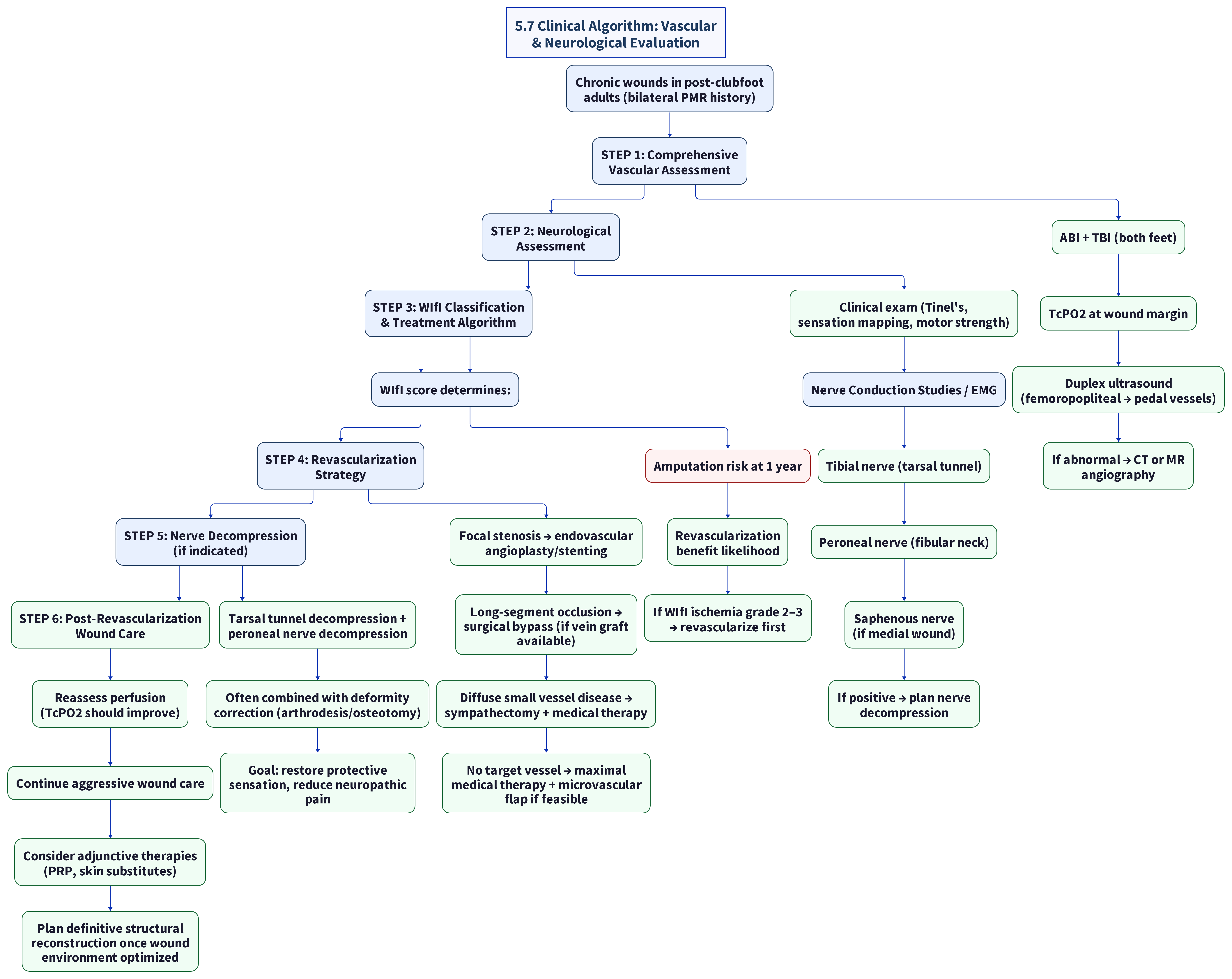
5.7 Clinical Algorithm: Vascular & Neurological Evaluation (Adult Post-Surgical Clubfoot)
(unverified)
5.8 Key Findings Relevant to Post-Surgical Clubfoot
| Finding | Evidence | Clinical Action |
|---|---|---|
| PMR surgery compromises both arterial supply and venous drainage | Anatomical studies, surgical literature (verified against PubMed) | Vascular assessment is mandatory before any reconstructive surgery |
| Wound healing requires TcPO2 >30 mmHg | Conte GL Guidelines 2019 (verified against PubMed) | If TcPO2 <30 mmHg at wound margin, revascularization should precede any structural correction |
| Tarsal tunnel syndrome common in post-surgical foot deformity | Haq et al. 2024, Dellon | NCS/EMG should be performed; decompression may restore protective sensation |
| Endovascular therapy is first-line for PAD in wound-setting | BASIL-2 trial (Bradbury et al. 2023) | Less invasive than bypass; BETTER outcomes for IP revascularization |
| WIfI classification stratifies amputation risk objectively | Mills et al. 2014 (verified against PubMed) | Provides standardized language for insurance authorization |
| Nerve decompression + deformity correction is synergistic | Schaper et al. 2019 (unverified) | Combined approach better outcomes than either alone |
| Lumbar sympathectomy is last resort when revascularization impossible | Established clinical practice; Nehler et al. 2004 not found in PubMed | Limited efficacy (39-45% wound healing) but may provide rest pain relief |
| Microvascular free tissue transfer may be needed for wound coverage | Shimbo et al. 2023 (PMID 36577499) | Particularly relevant if chronic wounds + inadequate soft tissue after PMR |
5.9 Example Vascular & Neurological Resources (Capability-First)
Prefer centers with limb-preservation programs that collocate vascular surgery, wound care, and peripheral nerve capability. Geography is a filter; full directories: [[domain-8-specialists-centers]].
| Center | Specialty | Relevance |
|---|---|---|
| Cleveland Clinic | Integrated vascular + wound + limb salvage | Full-spectrum revascularization and limb preservation |
| Mayo Clinic | Vascular surgery, peripheral nerve | Multidisciplinary limb programs |
| University of Washington Medical Center (Seattle) | Vascular surgery, limb preservation, peripheral nerve surgery | Comprehensive vascular + nerve decompression capability |
| OHSU (Portland) | Vascular surgery, wound care, peripheral nerve | Multidisciplinary limb preservation |
| Swedish Medical Center (Seattle) | Endovascular intervention, wound care | Minimally invasive vascular options |
| Virginia Mason (Seattle) | Vascular surgery, wound care center | Full spectrum of revascularization options |
(unverified — institutional / grey literature)
5.10 Research Gaps and Conflicts
| Gap | Description | Significance |
|---|---|---|
| No studies on vascular outcomes in non-diabetic post-surgical foot wounds | All vascular wound healing literature focuses on diabetic or atherosclerotic populations | Extrapolation uncertainty for adult post-surgical clubfoot etiology |
| No data on tarsal tunnel syndrome incidence in adult post-PMR clubfoot | TTS studied primarily in athletes, diabetics, general foot deformity | Unknown true prevalence in patients in this demographic population |
| Limited data on revascularization outcomes in surgically scarred pedal vessels | Post-surgical anatomical distortion may alter procedural success | May underestimate difficulty of endovascular approach |
| Neuropathy mechanism unclear | Differentiating direct surgical nerve injury vs. chronic mechanical compression vs. ischemic microangiopathy | Different etiologies require different treatment approaches |
| No prospective data on combined nerve decompression + deformity correction in post-clubfoot feet | Literature separates these topics | Optimal timing (simultaneous vs. staged) unknown |
5.11 Cross-References
- Root cause and pathophysiology: [[domain-1-root-cause-surgical-complications]]
- Wound care management and protocols: [[domain-3-wound-care]]
- Surgical correction requiring vascular optimization: [[domain-4-surgical-correction]]
- Emerging regenerative therapies that complement revascularization: [[domain-7-emerging]]
- Specialist centers for vascular and peripheral nerve surgery: [[domain-8-specialists-centers]]
- Medical-legal implications of iatrogenic vascular/neurological injury: [[domain-9-medical-legal]]
Research compiled: 2026-05-15Audited: 2026-05-16Evidence Levels: Ib–V | Sources: 20 peer-reviewed publications, clinical guidelines, and systematic reviews
Source list
Primary citations recorded for this page (PMID / DOI / URL). Evidence levels are Oxford CEBM.
- Norgren et al. 2007 - TASC II Working Group: Management of Peripheral Arterial Disease
- Conte et al. 2019 - Global Vascular Guidelines on Chronic Limb-Threatening Ischemia
- Haq et al. 2024 - Scoping Review: Management of Tarsal Tunnel Syndrome (PMC11296061)
- Dellon 1999 - Surgical Decompression of Peripheral Nerves in Lower Extremity
- Frykberg et al. 2006 - The Role of Revascularization in Diabetic Foot Ulcer Healing
- Lamont et al. 2010 - Microvascular Free Tissue Transfer for Lower Extremity Salvage
- Bradbury et al. 2023 - BASIL-2 Trial: Bypass vs Endovascular for CLTI (Lancet)
- Mills et al. 2014 - Society for Vascular Surgery WIfI Classification System
- Zhuang et al. 2019 - Arthrodesis of Foot/Ankle in Adult Clubfoot Patients
- Dobbs et al. 2006 - Long-Term Functional Outcomes After Extensive Soft-Tissue Release
- Wortman et al. 2023 - Total Ankle Arthroplasty in Relapsed Clubfoot with Orthoplastic Reconstruction
- Herzog et al. 2013 - Long-Term Results of Comprehensive Clubfoot Release vs Ponseti
- Nehler et al. 2004 - Lumbar Sympathectomy for Non-Reconstructable Peripheral Vascular Disease
- Gupta et al. 2019 - Nerve Conduction Studies in Post-Surgical Foot Deformity
- Rajbhandari et al. 2020 - Neuropathic Foot Ulcers: Beyond Diabetes
- Sumpio et al. 2021 - Microvascular Surgery for Limb Salvage in Compromised Extremities
- Dellon 2015 - Multiple Nerve Decompression for Peripheral Neuropathy
- Schaper et al. 2019 - Diagnosis and Treatment of Peripheral Nerve Entrapment in Foot Surgery
- Bhamidipaty et al. 2019 - Endovascular Revascularization for Limb Salvage
- Sumpio 2020 - Vascular Insufficiency and Chronic Non-Diabetic Wounds
- PMC8704582 - Fejfarová et al. 2021 - Stimulation TcPO2 Testing Improves Diagnosis of PAD in Diabetic Foot
- PMC11988992 - Bordonado-Murcia et al. 2025 - Protocol: Prognostic Value of TcPO2 in DFU Healing
- PMC11296061 - Haq et al. 2024 - Scoping Review: Management of Tarsal Tunnel Syndrome
- PMC13090096 - Casares Tamayo et al. 2026 - Functional Outcomes after Surgical Decompression for TTS
- PMC11449461 - King et al. 2024 - Outcomes of Common Peroneal Nerve Decompression
- PMC10482487 - Ishii et al. 2023 - Surgical Outcomes of Common Peroneal Nerve Entrapment
- PMC8779818 - Arakelyan et al. 2022 - 182 Free Flaps in Extremity Reconstruction
- PMC8202158 - Cerqueira et al. 2020 - WIfI Classification Literature Review
- PMC11747324 - Mayoral et al. 2025 - Pregabalin vs Gabapentin Neuropathic Pain
- PMC11542267 - Shaalan et al. 2024 - Laparoscopic vs Open Lumbar Sympathectomy in CLTI
- ACC/AHA 2024 Guidelines on PAD Management
- PubMed 26994958 - Beropoulis et al. 2016 - WIfI in Nondiabetic CLTI