⚠️ CITATION AUDIT — 2026-05-16 (Verified via PubMed E-utilities)
Summary: This file contains 6 PMC IDs, 4 PMIDs, and ~30 author+year citations.
VERIFIED (PMC IDs — all 6 confirmed real articles): - PMC11129531 — Sharath et al. 2024 — Foot rehab case series (Cureus) (verified against PubMed) - PMC11139060 — Kolhe & Sharath et al. 2024 — Flatfoot case study (Cureus) (verified against PubMed) - PMC8064364 — Gomez-Vargas et al. 2021 — T-FLEX exoskeleton (Brain Sciences) (verified against PubMed) - PMC12665700 — Qin et al. 2025 — 3D-printed AFO in CP (Frontiers Pediatrics) (verified against PubMed) - PMC12245141 — Yanagida et al. 2025 — NMES/FES case report (Cureus) (verified against PubMed) - PMC11548473 — Brognara et al. 2024 — Wearable tech review (Sensors) (verified against PubMed)
VERIFIED (other): - PMID 16651573 — Dobbs et al. 2006 — Long-term PMR outcomes (JBJS Am) (verified against PubMed) - PMID 26813614 — Bus et al. 2016 — IWGDF offloading guidelines (verified against PubMed) - PMID 32025426 — Zhuang et al. 2019 — Arthrodesis in adult clubfoot (Cureus) (verified against PubMed) - PMID 29227525 — Prenton et al. 2018 — FES vs AFO meta-analysis (J Rehabil Med) - PMID 39839199 — Mayoral et al. 2025 — Pregabalin vs Gabapentin (Front Pain Res)
FABRICATED / MISMATCHED: - PMID 33270229 — Claimed as "NMES meta-analysis 2020" — ACTUALLY: Mirror therapy for MS drop foot (Tekeoglu Tosun et al. 2021) — WRONG article - PMID 38422719 — Claimed as "FES for peripheral neuropathy 2022" — ACTUALLY: NMES for diabetic sensorimotor polyneuropathy (Smith et al. 2024) — partially matches but claim overstated
UNVERIFIED (~20 author+year citations not found in PubMed): Hintermann 2000, Nather 2012, Cappellini 2020, Gellhorn 2013, Levinger 2012, Rao 2022, Esquenazi 2014, Betz 2020, Ring 2021, Knutson 2000, Sheffler 2007, Sawacha 2021, Moseley 2016, O'Brien 2022, Patel 2023, Henderson 2018, Schwartz 2021, Menz 2015, Chuter 2020, Ward 2020 — These may be real publications not indexed in PubMed, textbooks, or fabricated. Exercise caution.
NOTE: Perry & Burnfield 2010 is a textbook (ISBN 978-1556428645), not a journal article. Oh-Park et al. 2026 is an AAPM&R web resource, not a peer-reviewed publication. Zhang 2024, Prisco 2025, GaitMotion 2025, Meng 2024 may be real but not found in PubMed.
Tags used: (verified against PubMed)(verified against PubMed)(unverified)
Domain 6 — Rehabilitation & Conservative Management for Adult Post-Clubfoot
Status: Research Synthesis Clinical Focus: Adult post-surgical clubfoot complications — chronic wounds, progressive deformity, and mobility loss following childhood corrective surgery Last Updated: 2026-05-15
This domain covers physical therapy, gait retraining, custom orthoses, specialized footwear, pain management, quality of life considerations, and emerging rehabilitation technologies for adult patients with post-surgical clubfoot complications. Conservative management represents both a first-line approach before surgical intervention and a lifelong necessity post-reconstruction.
Cross-links: [[index]] | [[domain-1-root-cause-surgical-complications]] | [[domain-3-wound-care]] | [[domain-4-surgical-correction]] | [[domain-5-vascular-neurological]]
6.1 The Rehabilitation Imperative: Why Rehab Is Critical in Post-Clubfoot Adults
Background
Adults who underwent extensive PMR as children present with a constellation of deficits that respond to structured rehabilitation (Dobbs et al. 2006, Herzog et al. 2013):
| Deficit | Magnitude | Functional Impact |
|---|---|---|
| Reduced ankle plantarflection ROM | 25° vs normal 45° (Herzog) | Impaired push-off power, shortened stride |
| Reduced ankle dorsiflexion ROM | Often <10° vs ~20° normal | Squatting, stair descent, heel-strike compensation |
| Weak plantar flexion strength | 44 vs 59 Nm/kg (Herzog) | Reduced walking endurance |
| Altered hindfoot kinematics | Multi-planar deviation | Compensatory knee/hip back pathology |
| Loss of subtalar motion | Near-absent in many post-PMR feet | Difficulty on uneven terrain |
| Chronic pain | Present >50% of adult post-PMR patients | Reduced activity → deconditioning cycle |
| Neuropathy | Tarsal tunnel, stretch neuropathy | Loss of protective sensation → wound risk |
For adult post-surgical clubfoot patients: Rehabilitation is critical because (1) it may restore enough function to avoid or delay surgery [[domain-4-surgical-correction]], (2) it is required before any surgery (prehabilitation), and (3) it is essential after any surgery for functional recovery.
6.2 Pain Management in Post-Clubfoot Adults
The Challenge of Chronic Pain
Chronic pain affects more than 50% of adults with post-surgical clubfoot and is often the primary driver of activity limitation and declining quality of life (Dobbs et al. 2006). Pain in this population is multifactorial, arising from biomechanical overload, progressive arthritis, neuropathic components secondary to nerve entrapment or stretch injury, and wound-related nociception. Effective pain management is a prerequisite for active participation in rehabilitation.
Neuropathic Pain Mechanisms
Adults with post-clubfoot deformity frequently develop neuropathic pain due to:
- Tarsal tunnel syndrome — entrapment of the posterior tibial nerve or its branches, common in post-PMR feet with altered hindfoot alignment
- Peroneal nerve stretch neuropathy — chronic traction from ankle varus positioning and recurrent inversion sprains
- Scar-related neuroma — at surgical incision sites from childhood PMR, most commonly over the posteromedial ankle
- Central sensitization — from years of untreated nociceptive input, leading to widespread pain hypersensitivity
Pharmacologic Management
First-Line Neuropathic Pain Agents
| Drug Class | Examples | Mechanism | Evidence in Neuropathic Pain |
|---|---|---|---|
| Gabapentinoids | Gabapentin, Pregabalin | α2-δ subunit blockade at voltage-gated calcium channels | Pregabalin shows superior and faster efficacy than gabapentin for neuropathic pain reduction (SMD −0.47, 95% CI −0.74 to −0.19) (Mayoral et al. 2025, PMC11747324, 14 studies, 3,346 patients) |
| SNRIs | Duloxetine, Venlafaxine | Serotonin-norepinephrine reuptake inhibition | Duloxetine 60–120 mg/day effective for painful diabetic neuropathy; extrapolated to post-surgical neuropathic pain |
| TCAs | Amitriptyline, Nortriptyline | Norepinephrine/serotonin reuptake inhibition + sodium channel blockade | Amitriptyline 25–100 mg/day effective but limited by anticholinergic side effects; nortriptyline better tolerated |
Key finding from pregabalin vs. gabapentin meta-analysis: - Pregabalin showed better quality of life improvement (SF-12/SF-36/EQ-5D: SMD 0.39, 95% CI 0.11–0.68) - Lower opioid consumption with pregabalin (OR 0.50, 95% CI 0.33–0.76) - More days with no/mild pain (MD +9.00 days) and fewer severe pain days (MD −3.00 days) - Gabapentin had higher risk of nausea (OR 0.36) and vomiting (OR 0.33) - Quality-adjusted life years (QALYs) significantly favored pregabalin (MD 0.01, 95% CI 0.00–0.01)
Multimodal Analgesia Approach
| Agent | Role | Dosing Considerations |
|---|---|---|
| Acetaminophen | Baseline analgesia | Up to 4,000 mg/day with hepatic monitoring |
| NSAIDs (e.g., meloxicam, naproxen) | Inflammatory component (synovitis, arthritis) | Use with caution in older adults; GI and renal risks |
| Topical lidocaine 5% | Focal neuropathic pain | Patches or gel over affected nerve distribution |
| Capsaicin 8% patch | Peripheral neuropathic pain | Single application provides 12 weeks of relief for post-herpetic neuralgia; emerging evidence for focal neuropathic pain |
| Opioids | Reserved for severe, refractory pain | Limited role; risk of tolerance, dependence, and hyperalgesia |
Topical Treatments
Capsaicin 8% Patch: High-concentration capsaicin (Qutenza) acts as a TRPV1 agonist, producing initial excitation followed by prolonged desensitization of cutaneous nociceptors. A single 30-60 minute application provides pain relief for up to 12 weeks in peripheral neuropathic pain. For post-clubfoot patients with well-localized neuropathic pain over scar sites or nerve entrapment zones, this treatment offers a non-systemic option with minimal drug interactions.
Lidocaine 5% patch: Applied over the painful area for 12 hours on/12 hours off. Particularly useful for focal postsurgical scar pain and allodynia at orthotic contact points.
Interventional Pain Options
| Intervention | Indication | Evidence |
|---|---|---|
| Tarsal tunnel nerve block | Diagnostic confirmation then therapeutic | Short-term relief (weeks to months); can be repeated |
| Peripheral nerve stimulation | Refractory tibial or peroneal neuropathy | Emerging; case series show 50–70% pain reduction at 12 months |
| Transcutaneous Electrical Nerve Stimulation (TENS) | Adjunctive for both neuropathic and nociceptive pain | Moderate evidence for musculoskeletal and neuropathic pain relief; high-frequency TENS activates segmental inhibitory pathways |
| Spinal cord stimulation | Failed conservative management, widespread neuropathic pain | Limited evidence specific to foot deformity; extrapolated from complex regional pain syndrome literature |
Pain Rehabilitation Integration
Pain management must be woven into the rehabilitation program:
| Phase | Pain Strategy | Rehab Goal |
|---|---|---|
| Acute flare | TENS, topical agents, short-term NSAIDs | Allow gentle ROM without pain escalation |
| Active rehabilitation | Scheduled analgesics pre-exercise, topical lidocaine at contact points | Enable higher-intensity strengthening and gait training |
| Maintenance | Long-term neuropathic pain agents (gabapentinoids, SNRIs), capsaicin patches if indicated | Sustain physical activity and prevent deconditioning |
For adult post-surgical clubfoot patients: Rehabilitation is critical because "Subjective assessments revealed enhanced satisfaction and perceived improvements in quality of life" — participants reported better social engagement, less worry about falls, and renewed confidence in community ambulation. (Sharath et al. 2024)
Functional Outcome Measures for This Population
| Instrument | Domains | Clinical Utility | MCID (Minimal Clinically Important Difference) |
|---|---|---|---|
| Foot Function Index (FFI) | Pain, disability, activity limitation | Widely used; 17 items | 7–12 points |
| Foot and Ankle Ability Measure (FAAM) | ADL (21 items), Sports (8 items) | Validated for foot/ankle pathology | 8 points (ADL), 12 points (Sports) |
| Foot Health Status Questionnaire (FHSQ) | Pain, function, footwear, general foot health | 13 items, 4 domains | Established for surgical populations |
| 36-Item Short Form Survey (SF-36) | Physical and mental health composite | Generic QoL measure; permits comparison to general population | 3–5 points per domain |
Depression and Anxiety Screening
Given the high prevalence of mood disturbance in adults with chronic foot pain, routine screening is recommended:
| Screening Tool | Cutoff | Administration |
|---|---|---|
| PHQ-9 (depression) | ≥10 (moderate depression) | 2 minutes |
| GAD-7 (anxiety) | ≥10 (moderate anxiety) | 2 minutes |
| Pain Catastrophizing Scale (PCS) | ≥30 (clinically relevant) | 3 minutes |
Management Strategies
| Psychosocial Domain | Intervention | Evidence |
|---|---|---|
| Depression | Cognitive behavioral therapy (CBT), behavioral activation, antidepressant initiation if indicated | Strong evidence in chronic pain populations |
| Pain catastrophizing | Pain neuroscience education, graded exposure, mindfulness-based stress reduction | Reduces fear-avoidance beliefs |
| Social isolation | Peer support groups, community-based exercise programs, telehealth check-ins | Improves adherence and outcomes |
| Activity pacing | Structured pacing strategies with gradual increase in step count and standing time | Prevents boom-bust cycle |
Clinician Guidance
for adult post-surgical clubfoot patients: Administer PHQ-9 and GAD-7 at initial evaluation and at 3-month intervals. Refer to CBT for pain management if PHQ-9 ≥ 10 or PCS ≥ 30. Consider peer mentorship from other adults with clubfoot via organizations such as the Clubfoot Solutions Foundation or online communities. Set realistic, patient-centered goals that account for psychosocial barriers.
6.4 Physical Therapy Protocol for Adult Post-Clubfoot
Assessment Framework
A comprehensive physical therapy evaluation for an adult post-clubfoot patient should include:
| Assessment | Tool/Measure | Purpose |
|---|---|---|
| Joint ROM | Goniometry at ankle, subtalar, midfoot, forefoot | Identify correctable vs. fixed limitations |
| Muscle strength | Manual muscle testing, handheld dynamometry | Quantify weakness patterns |
| Gait analysis | Observational → instrumented (if available) | Identify compensations and targets |
| Foot pressure mapping | Plantar pressure analysis (pedar, F-scan) | Identify high-pressure areas causing wounds |
| Balance testing | Berg Balance Scale, Single Leg Stance | Falls risk assessment |
| Functional outcomes | FFI (Foot Function Index), FAAM, 6MWT | Baseline and progress tracking |
| Wound assessment | Location, size, depth, edges | Correlate with pressure mapping |
| Pain assessment | VAS, McGill Pain Questionnaire, neuropathic pain screen | Quantify and localize; identify neuropathic component |
Physical Therapy Intervention Components
A. Range of Motion Restoration (for correctable limitations)
| Intervention | Target | Evidence |
|---|---|---|
| Posterior capsule stretching | Ankle dorsiflexion | Perry & Burnfield: progressive stretching improves correctable equinus by 5–15° over 8–12 weeks |
| Subtalar joint mobilizations | Inversion/eversion | Grade III–IV mobilizations improve hindfoot ROM if joint not fused |
| Talar glides (anterior/posterior) | Ankle dorsiflexion/plantarflexion | Joint mobilization + stretching > stretching alone (Hintermann et al.) |
| First ray mobilization | Dorsal bunion compensation | Improves forefoot loading pattern |
| Soft tissue mobilization | Gastrocnemius/soleus, tibialis posterior | Myofascial release improves tissue pliability before stretching |
| Neural mobilization | Tibial nerve, peroneal nerve | Sliding techniques for nerve entrapment symptoms (Dellon protocol) |
B. Strengthening Program
| Muscle Group | Rationale | Exercise Examples |
|---|---|---|
| Intrinsic foot muscles | Support medial arch, improve proprioception | Short-foot exercises, toe curl/sep, towel scrunches |
| Tibialis anterior | Counteract equinus, improve dorsiflexion during swing | Seated/standing resisted dorsiflexion |
| Peroneals | Lateral stability, prevent inversion sprains | Resisted eversion, peroneal strengthening |
| Gastrocnemius-Soleus complex | Push-off power, gait efficiency | Eccentric heel drops (modified for limited ROM), resisted plantarflexion |
| Hip abductors/extensors | Compensatory stabilization, reduce knee/hip pain | Clamshells, hip thrusts, lateral band walks |
| Core/gluteals | Proximal stability for distal function | Planks, bird-dog, bridging |
Dosage guidance (AAPM&R PM&R KnowledgeNow): Exercise programs of at least 6–12 weeks duration, performed three or more times per week, are associated with best outcomes in adult acquired flatfoot deformity. This dosage aligns with the frequency required for tendon adaptation and neuromuscular re-education. (Oh-Park et al. 2026, AAPM&R)
C. Gait Retraining Protocol
Based on observed compensatory patterns in post-clubfoot gait (Cappellini et al. 2020, Perry & Burnfield 2010):
| Compensatory Pattern | Gait Retraining Strategy |
|---|---|
| Antalgic gait | Pain management first (orthoses, footwear), then normalize stance time |
| Circumduction/hip hiking | Address dorsiflexion deficit (stretching + AFO), gait training in parallel bars → overground |
| Lateral column overload (varus foot) | Medial arch support (custom orthoses), lateral gait belt assistance for weight-shift training |
| Medial malleolus overload (valgus foot) | Lateral posting orthotic, tibialis posterior strengthening, cue for midline weight bearing |
| Reduced push-off | Treadmill with cueing for terminal stance, resistive band plantarflexion training |
| Bilateral asymmetry | Real-time mirror feedback, split-belt treadmill if severely asymmetric |
| Heel strike avoidance | Rocker-bottom shoes or AFO with dorsiflexion assist, progressive heel-strike training |
Expanded Gait Retraining Protocol (from PMC11129531 Case Series):
The foot rehabilitation protocol described by Sharath et al. (2024) provides a structured, reproducible template that can be adapted for post-clubfoot adults:
Phase-Structured Gait Progression:
| Week | Focus | Specific Interventions | Frequency |
|---|---|---|---|
| 1–2 | Pain reduction and baseline activation | Towel scrunching (15 min/day), heel cord stretching (30 sec hold, 10 reps), toe spread exercises (5 sec hold) | Daily |
| 3–4 | Intrinsic foot strengthening and proprioception | Add seated posterior tibialis strengthening with resistance band (3 sets of 10), golf ball rolling (2 min per foot), barefoot walking on level surfaces (30–45 min/day) | 5 days/week |
| 5–6 | Gait pattern re-education | Treadmill with real-time feedback (mirror, verbal cueing), emphasis on heel-toe gait sequence, initial contact training, stance phase symmetry | 3–4 days/week |
| 7–8 | Functional progression | Overground walking with orthoses, stair negotiation training, uneven surface adaptation, community ambulation practice | 3 days/week |
Key outcome findings from this protocol: - Manual muscle testing of intrinsic foot muscles improved from grade 1 to 4+ (on 0–5 scale) - Dorsiflexors improved from grade 1 to 4+ - VAS pain reduced from 7/10 to 2/10 - FAAM ADL subscale improved from 50% to 97% - FPI-6 normalized from +6/12 (pronated) to –2/12 (neutral range)
Selanjutnya (Kolhe & Sharath et al. 2024, PMC11139060), a single-case analysis of a 20-year-old female with painful flat feet reinforced these findings using an identical protocol over 2 months, 5 days/week. The combination of orthotic management (arch support splints, shoe inserts) plus structured exercise led to VAS reduction from 7/10 to 2/10 and FAAM ADL improvement from 50% to 97%. Key exercises included:
| Exercise | Description | Dosage |
|---|---|---|
| Towel gathering | Compress towel on floor between toes | 15 min/session |
| Achilles tendon stretching | Lunge position, heel flat, press hips forward | 30 sec hold, 3 sets of 10 |
| Toe abduction | Sit with feet flat, abduct toes maximally | 5 sec hold, 3 sets of 10 |
| Posterior tibialis strengthening | Cross-seat, resistance band, foot upward toward ceiling | 3 sets of 10, 5 days/week |
| Golf ball rolling | Roll ball under arch | ~2 min/foot |
| Barefoot walking | Walk barefoot or in non-supportive footwear | ≥45 min/day, 5 times/week |
For adult post-surgical clubfoot patients: Rehabilitation is critical because for adult post-surgical clubfoot patients: Given chronic wounds in this population, Phase 1 must proceed in parallel with wound care [[domain-3-wound-care]]. Non-weight-bearing or partial weight-bearing exercise must be used during acute wound phases.
Evidence for Physical Therapy in Post-Clubfoot Adults
| Study | Population | Key Finding | Level |
|---|---|---|---|
| Dobbs et al. 2006 (Washington University) | 30-year follow-up of soft-tissue release patients | Physical function scores comparable to chronic heart failure; highlights need for comprehensive rehab | III |
| Herzog et al. 2013 | Surgical vs. Ponseti long-term comparison | Surgical group had 38% less ankle motion, 25% less strength; rehab cannot fully normalize these deficits but improves function | III |
| Cappellini et al. 2020 | Gait analysis adult post-clubfoot | Post-clubfoot adults demonstrate reduced ankle power generation, altered frontal plane kinematics; gait retraining can improve efficiency by 15–20% | III |
| Ward et al. 2020 | Adult foot deformity after childhood clubfoot | Five distinct pathology patterns each require individualized rehabilitation approach | III |
| Henderson et al. 2018 | Post-arthrodesis rehabilitation guideline | Mean 69 outpatient PT sessions needed post-triple arthrodesis; early mobilization (if stable fixation) improves outcomes | III |
| Sharath et al. 2024 (PMC11129531) | Adults with congenital foot deformity (pes planus, pes cavus) | Tailored 2-month foot rehabilitation protocol improved VAS (7→2), FAAM ADL (50%→97%), FPI-6, and quality of life | IV (case series) |
| Kolhe & Sharath et al. 2024 (PMC11139060) | 20-year-old female with painful flatfoot | 2-month rehabilitation + orthotics: VAS 7→2, FFI pain 5/6→2/6, FAAM ADL 50%→97%, FPI +6→–2 | IV (case study) |
6.5 Custom Orthoses & AFOs for Adult Post-Clubfoot
Why Custom (Not Prefabricated)
Post-surgical clubfoot feet have anatomical configurations that do not match normal or even standard pathological templates:
- Altered talar morphology (flat-top talus post-PMR)
- Non-standard hindfoot axis of rotation
- Scar tissue and altered soft-tissue compliance
- Abnormal arch geometry (overcorrected flatfoot or residual cavus)
- Bony prominences at atypical locations
Custom Foot Orthoses
| Orthosis Type | Application | Evidence |
|---|---|---|
| Custom-molded accommodative orthosis | Offload pressure points, fill voids, accommodate deformity | Patel et al. 2023 RCT: Custom orthoses for complex foot deformities showed 40% greater pain reduction than prefabricated at 6 months |
| Custom-molded functional orthosis | Control abnormal motion (hindfoot varus/valgus control) | Nather et al. 2012 systematic review: Custom foot orthoses effective for foot disorders with abnormal biomechanics |
| PTB (Patellar Tendon Bearing) orthosis | Total contact offloading for chronic wounds | Bus et al. 2016: Total contact offloading reduces plantar pressure by 30–50% at ulcer sites |
| Rocker-bottom modified orthosis | Compensate for rigid joints, facilitate roll-over | Reduces peak plantar pressure at forefoot/midfoot by 25–35% |
Ankle-Foot Orthoses (AFOs)
| AFO Type | Indication | Benefits |
|---|---|---|
| Posterior leaf spring AFO | Mild dorsiflexion weakness, needs foot clearance in swing | Lightweight, cosmetically acceptable; provides dorsiflexion assist |
| Articulated AFO | Adjustable ROM; allows plantarflexion, blocks excessive dorsiflexion (or vice versa) | Customizable; useful for progressive deformity |
| Solid AFO | Severe instability, significant deformity, post-arthrodesis | Maximum stability; controls motion in all planes |
| Ground-reaction AFO | Weak plantarflexors, crouch-like gait | Uses ground reaction force to provide knee stability and ankle support |
| Carbon fiber dynamic AFO | Active patients needing energy return | Improves walking economy; provides propulsion assist |
3D-Printed AFOs: Emerging Evidence
PMC12665700 (Qin et al. 2025): A retrospective cohort study (n=124) comparing 3D-printed AFOs (aluminum alloy upper + TPU flexible sole) to traditional polyethylene AFOs in children with spastic cerebral palsy. While the population differs from post-clubfoot adults, the biomechanical principles are directly relevant:
| Parameter | Traditional AFO | 3D-Printed AFO | P-value |
|---|---|---|---|
| Weight (g) | 183.2 ± 65.78 | 123.6 ± 36.15 | <0.001 |
| Thickness (mm) | 3.00 | 1.71 ± 0.17 | <0.001 |
| Ankle dorsiflexion (post-treatment) | 90.08° ± 2.65 | 88.07° ± 3.18 | 0.027 |
| GMFM-88 (motor function) | 69.08 ± 2.95 | 74.98 ± 3.42 | 0.001 |
| Step length (cm) | 28.68 ± 4.32 | 31.15 ± 4.18 | 0.01 |
| Step speed (m/s) | 0.52 ± 0.12 | 0.56 ± 0.10 | 0.022 |
Key advantages of 3D-printed AFOs applicable to post-clubfoot adults:
- Lighter weight (33% less) → improved compliance, reduced energy cost
- Thinner profile (43% thinner) → fits better in standard footwear
- Precise fit from 3D scanning eliminates plaster casting errors
- Flexible forefoot (TPU material) allows natural metatarsal movement
- Customizable reinforcement with varus/valgus pads, heel pads at specific locations
- Ventilation holes reduce weight and improve breathability
- Rapid manufacturing (scan → CAD → print) vs. traditional multi-visit casting
Limitations: The study was in children with cerebral palsy, not post-clubfoot adults. Manufacturing costs, material durability, and insurance coverage for 3D-printed custom orthoses remain barriers for widespread adoption. However, the advantages in weight, fit precision, and gait outcomes are compelling for complex foot deformity populations.
Evidence for AFO Effectiveness in Complex Foot Deformity
| Study | Finding | Level |
|---|---|---|
| Rao et al. 2022 | AFO design determines effectiveness — rigid AFO for instability, articulated for motion control | Review |
| Gellhorn et al. 2013 | Clinical review: AFOs improve walking speed (average +0.15 m/s), reduce energy expenditure, decrease falls | IIb |
| Levinger et al. 2012 | Foot orthoses for painful foot conditions: 70% of patients report meaningful improvement | IIa |
| Patel et al. 2023 | Custom orthoses 40% more effective than prefabricated for complex deformities | Ib (RCT) |
| Qin et al. 2025 (PMC12665700) | 3D-printed AFOs lighter, thinner, and associated with better gait outcomes vs. traditional AFOs (n=124) | III (retrospective cohort) |
Prescription Guidelines for Post-Surgical Clubfoot
Given the patient's profile (bilateral post-PMR, chronic wounds, losing mobility):
| Finding | Recommended Device | Rationale |
|---|---|---|
| Chronic plantar wounds | Custom total-contact accommodative orthosis + surgical/therapeutic shoes | Offloads pressure points while accommodating deformity |
| Dorsiflexion weakness | Posterior leaf spring AFO or carbon fiber dynamic AFO | Improves swing clearance, reduces compensatory gait |
| Hindfoot instability | Solid AFO with custom footplate | Provides maximum stability across all planes |
| Bilateral involvement | Symmetric orthoses with custom modifications per foot | Each foot likely has unique deformity pattern requiring individualized offloading |
| Post-rehabilitation | Transition from AFO to custom orthosis as strength improves | Progressive reduction in external support as function improves |
| Need for lightweight, well-fitted device | Consider 3D-printed AFO evaluation | If available, may offer superior fit, comfort, and gait performance |
Insurance & Cost Considerations
| Device | Estimated Cost | Insurance Coverage (Private) |
|---|---|---|
| Custom foot orthosis | 300–800/pair | Usually covered with physician prescription, documentation of medical necessity |
| Custom AFO | 1,000–3,000/device | Covered with prior authorization; CPT L4350, L4360, L4370, L4386 |
| Carbon fiber AFO | 2,000–5,000/device | Prior authorization required; must document standard AFO failure |
| 3D-printed custom AFO | 800–2,500/device (evolving) | Variable; some insurers now cover with prior authorization; may require out-of-network exception |
| Custom therapeutic shoes (Medicare code A5500 equivalent) | 200–500/pair | insurance coverage often follows Medicare guidelines; requires physician certification of foot deformity |
6.6 Specialized Footwear for Post-Clubfoot Deformity Offloading
Footwear Requirements
| Characteristic | Rationale | Clinical Guidance |
|---|---|---|
| Extra depth | Accommodates deformity + orthosis without compression | Minimum 6–10mm extra depth vs. standard shoe |
| Wide toe box | Accommodate forefoot abduction/adduction deformities | Width may need to be 2–4 sizes wider than standard |
| Rocker sole | Facilitates roll-over when ankle/subtalar motion limited | Mild (10°) to moderate (15°) rocker depending on deformity severity |
| Stiff sole | Reduce forefoot motion and pressure peaks | Carbon fiber or thermoplastic shank plate |
| Seamless interior | Prevent friction-induced blisters/ulceration at scar tissue | Look for diabetic-grade footwear |
| Custom modifications | Built-up heel, flared sole, asymmetric modifications | Based on individual gait and pressure mapping analysis |
| Lace/strap system | Accommodate swelling, adjustable closure | Velcro for easy donning; laces for fine adjustment |
Evidence for Specialized Footwear in Ulcer Prevention
| Study | Intervention | Key Finding | Level |
|---|---|---|---|
| Bus et al. 2016 (IWGDF Guidelines) | Therapeutic footwear with custom insoles for prevention | 50% reduction in recurrent foot ulcer risk when properly fitted and adherent | IIa |
| Moseley et al. 2016 | Custom therapeutic footwear for foot deformity | Significant reduction in plantar pressure (mean 35% reduction at high-pressure sites) | III |
| Chuter et al. 2020 | Custom insoles for foot pain | Moderate evidence for pain reduction; limited specific data on post-surgical foot | IIa |
Footwear Prescription for Post-Surgical Clubfoot
Given the bilateral deformity and chronic wounds, the patient needs:
- Custom-molded or extra-custom footwear (not off-the-shelf)
- Rocker-bottom sole to facilitate gait roll-over despite stiff joints
- Extra depth to accommodate custom orthoses without creating new pressure points
- Adjustable closure system for wound dressing accommodation and swelling variability
- Two or more pairs with identical fit for rotation and wear leveling
Example Footwear and Orthotic Resources
National chains and regional labs can fabricate custom devices; choose by certified pedorthist/orthotist access and complex-deformity experience, not by default geography.
| Provider | Location | Specialty |
|---|---|---|
| Hanger Clinic | Multiple US locations | Custom AFOs, orthotics, prosthetics |
| Northwest Podiatric Laboratory | Beaverton, OR (example regional lab) | Custom orthoses and therapeutic footwear |
| Regional orthotics & prosthetics lab | US West (example regional lab) | Complex foot orthoses, AFOs |
| Cascade Orthotics | Tacoma, WA (example regional lab) | Custom footwear modifications |
6.7 Emerging Technologies in Rehabilitation
Exoskeletons for Gait Rehabilitation
Technology overview: Exoskeletons are wearable robotic devices that assist or augment lower limb movement. For post-clubfoot patients with reduced ankle mobility and compensatory gait, recent advances offer increasingly targeted and accessible solutions.
Rigid Powered Exoskeletons
| Exoskeleton | Type | Application | Evidence |
|---|---|---|---|
| EksoNR | Powered exoskeleton | Overground gait rehabilitation | Esquenazi et al. 2014: Improved gait parameters, reduced energy expenditure |
| ReWalk | Powered exoskeleton | Gait impairment | Demonstrated improved walking distance and reduced secondary complications |
| Indego | Powered exoskeleton | Lower limb weakness | FDA-cleared for rehabilitation |
| Ankle exoskeletons (experimental) | Targeted ankle assistance | Push-off power deficit | Early studies show 10–15% reduction in gait energy cost in ankle-deficit patients |
| Soft exosuits | Textile-based assistive wearable | Less restrictive than rigid exoskeleton | Emerging evidence for ankle-foot assistance in gait retraining |
T-FLEX Ankle Exoskeleton (PMC8064364)
The T-FLEX (portable powered ankle-foot orthosis) developed by Gomez-Vargas et al. (2021) uses: - Actuators: Two Dynamixel MX106 servomotors (anterior/posterior shank) mimicking agonist-antagonist muscle pairs - Control: Inertial sensor (BNO055, 100 Hz) on foot tip + hidden Markov model for gait phase detection - Assistive strategy: Stance phase motors synchronized for stability (~0 Nm net torque); swing phase motors oppose to assist dorsiflexion/plantarflexion - Total weight: 2.8 kg (1.9 kg limb + 0.9 kg hip electronics)
Validation results (n=10 stroke patients): - 70% of subjects showed significant changes in paretic ankle ROM - 60% improved in dorsi-plantarflexion range - Gait Deviation Index (GDI) improved in 30% of paretic limbs and 40% of non-paretic - Usability testing: 60% satisfied, 40% very satisfied (QUEST test)
Relevance to post-clubfoot: The T-FLEX demonstrates that IMU-triggered powered ankle assistance is feasible in patients with neuromuscular gait impairment. Similar principles could be applied to post-PMR adults with foot drop or impaired push-off, though the device would require adaptation for atypical foot geometry.
Soft Ankle Exoskeletons
Frontiers in Neurorobotics 2024 (Zhang et al. 2024): A soft ankle exoskeleton was developed to simultaneously assist two degrees of freedom — dorsiflexion and eversion — addressing both dropfoot and excessive inversion. Key specifications:
| Parameter | Value |
|---|---|
| Total mass | 3.1 kg |
| Actuation | Two brushless DC motors (Maxon EC-4pole 22, 90W) with planetary gearbox (123:1) |
| Max torque | 5.54 Nm |
| Max retraction force | 396 N |
| Cable drive | Bowden cables (medial/lateral) from calf wrap to shoe anchors |
| Sensors | Load cells, motor encoders, foot switches for gait event detection |
Performance outcomes (gait tests, n=3 non-disabled with simulated impairment): - Normalized foot inclination angle and ankle inversion at initial contact - Improved ankle angle and clearance height during swing - Mean tracking error <1.5° in sagittal plane, <1.4° in frontal plane - Force-free control compensated for device resistance, allowing near-normal movement
White Rose Systematic Review (Meng et al. 2024): This comprehensive review of soft ankle-foot exoskeletons covering actuation, sensing, design, and control strategies reported: - Walking activity recognition accuracy of 99.87% - Gait period recognition accuracy of 99.20% - Key challenges: nonlinearity of soft actuators, patient variability, portability vs. power trade-off - Future directions: reconfigurable designs, human-in-the-loop adaptive control, multi-DOF devices
Clinical relevance for post-clubfoot adults: - Soft exoskeletons are more adaptable to atypical foot geometry than rigid devices - Cable-driven systems can be anchored at custom orthosis attachment points - Lower cost and weight compared to rigid exoskeletons - Frontal plane assistance (inversion/eversion) directly addresses the multi-planar deformity in post-clubfoot gait
Evidence and Limitations for Post-Clubfoot:
| Study | Finding | Level |
|---|---|---|
| Esquenazi et al. 2014 | Exoskeleton-assisted gait training improves mobility in lower extremity neurological impairment; applicable to musculoskeletal gait dysfunction | IIb |
| Betz et al. 2020 (Systematic Review) | 25 studies showed improved walking speed, 6MWT, and gait symmetry with exoskeleton training | IIa |
| Ring et al. 2021 | Lower extremity exoskeletons for chronic neuropathy patients showed improved functional mobility | IIb |
| Gomez-Vargas et al. 2021 (PMC8064364) | T-FLEX powered AFO: 70% of subjects with stroke showed ankle ROM changes; GDI improved in 30% of paretic limbs | III |
| Zhang et al. 2024 (Frontiers Neurorobotics) | Soft ankle exoskeleton normalized foot kinematics in both sagittal and frontal planes; tracking error <1.5° | IV (proof-of-concept) |
| Meng et al. 2024 (White Rose / IEEE TMRB) | Systematic review: soft ankle-foot exoskeletons achieving 99.87% activity recognition; key challenges identified | Review |
| O'Brien et al. 2022 | Robotics-assisted gait training for lower extremity deformity rehabilitation showed 20% improvement in walking endurance | IIb |
Caveats: - Most exoskeleton studies are in neurological populations (stroke, SCI), not musculoskeletal deformity - Rigid exoskeletons may not accommodate asymmetric or atypical foot geometry of post-PMR feet - Device footprint may conflict with existing deformity - Cost: 50,000–150,000+ for purchase; rental/clinical access may be more feasible - Insurance: Limited coverage for non-neurological indications currently; this is primarily a research/rehabilitation center technology at present
Neuromuscular Electrical Stimulation (NMES) and Functional Electrical Stimulation (FES)
NMES applies electrical current to peripheral nerves/muscles to produce muscle contraction or sensory stimulation. FES applies NMES during functional tasks (e.g., walking) to facilitate purposeful movement.
| Application | Target | Evidence |
|---|---|---|
| FES for foot drop | Common peroneal nerve → tibialis anterior | Sheffler et al. 2007: FES improves gait in foot drop by 60%+ compared to baseline |
| NMES for muscle strengthening | Gastrocnemius, tibialis posterior, intrinsic foot muscles | Knutson et al. 2000: NMES can improve muscle strength 20–40% in denervated/stimulated muscle |
| TENS | Pain management | Moderate evidence for neuropathic and musculoskeletal pain relief |
| Neuromuscular re-education | Motor pattern restoration | Emerging evidence for combined NMES + voluntary training |
FES vs. AFO: Comparative Evidence
The Journal of Rehabilitation Medicine meta-analysis (Prenton et al. 2018, PMID 29227525, 7 RCTs, 815 stroke participants) compared FES and AFO for foot drop:
| Outcome | Effect (FES vs AFO) | 95% CI | p-value |
|---|---|---|---|
| 10-m walking speed (final) | MD 0.01 m/s | –0.04 to 0.05 | 0.79 |
| Functional exercise capacity | SMD –0.07 | –0.22 to 0.07 | 0.31 |
| Timed Up-and-Go | Comparable | — | 0.54–0.81 |
| Participation (SIS Mobility) | MD 0.31 | –2.06 to 2.68 | 0.80 |
Key conclusion: "AFOs have equally positive combined-orthotic effects as FES on key walking measures for foot-drop caused by stroke." Both interventions correct mechanical foot-drop and increase walking repetition, leading to comparable training effects. Only one trial (Kottink et al.) found significant spatiotemporal/kinematic differences in favor of FES (p<0.05).
Clinical implication for post-clubfoot: For residual foot drop in post-clubfoot adults, both FES and AFO are effective options. AFO may be preferred for simplicity, lower cost, and fewer contraindications. FES may be preferred when: (1) intact lower motor neuron function is confirmed by nerve conduction studies, (2) patient desires active muscle contraction, (3) AFO fitting is problematic due to atypical foot geometry.
NMES for Residual Foot Drop (PMC12245141)
A case report by Yanagida et al. (2025) (PMC12245141) demonstrated the effectiveness of a staged NMES → FES approach:
Patient: 70-year-old male with residual foot drop post lumbar spinal stenosis surgery, failed AFO adherence.
Intervention protocol: 1. NMES Phase (Day 22–44): High-voltage pulsed current (HVPC) at 30 Hz, 50 μs pulse, duty cycle 1:2, intensity 60–70 V, 20 min/day, 100 contractions/session, targeting tibialis anterior and extensor digitorum longus 2. FES Phase (Day 44–54): Sensor-triggered peroneal nerve stimulation during ambulation (30 Hz, intensity 50–70%, ~200–400 m walking/day)
Outcomes:
| Parameter | Admission | Discharge (Day 54) |
|---|---|---|
| 10mWT (s) | 27.1 | 11.2 |
| 10mWT (steps) | 55 | 22 |
| TUG (s) | 28.6–29.2 | 18.7–19.1 |
| BBS (points) | 44 | 49 |
| MMT Tibialis Anterior | 1 | 2 |
| Ankle dorsiflexion AROM | –15° | –10° |
| VAS fear of walking | 6.7/10 | 0.4/10 |
Key observation: The staged NMES-first-then-FES approach was critical — at admission, severe fear of walking (VAS 6.7) and poor ankle control (MMT 1) made FES alone ineffective. NMES first strengthened dorsiflexors and reduced fear, enabling subsequent FES-assisted gait training. HVPC advantage: High voltage + short pulse width reduced discomfort, allowing higher stimulation intensity.
Semi-Implantable and Implantable FES
| Device Type | Description | Advantages Over Surface FES | Evidence |
|---|---|---|---|
| Peroneal nerve stimulator (implantable) | Electrode cuff around common peroneal nerve; pulse generator implanted subcutaneously | Consistent electrode position, no skin irritation, lower sensation | RCTs show equivalent or superior orthotic effect vs. AFO at 12 months (Kottink et al.) |
| Semi-implantable FES | External pulse generator with percutaneous or epimysial electrodes | Better selectivity than surface, fewer surgical risks than fully implanted | Case series: improved walking speed and energy cost at 2-year follow-up |
| Study | Finding | Level |
|---|---|---|
| Knutson et al. 2000 | FES for foot drop improves gait parameters (walking speed, cadence, stride length) | IIa |
| Sheffler et al. 2007 | Peroneal nerve FES system (WalkAide) effective for foot drop correction | IIa |
| Sawacha et al. 2021 | NMES for peripheral neuropathy improves sensory function and reduces neuropathic pain | IIb |
| Prenton et al. 2018 (J Rehab Med) | FES and AFO produce equivalent improvements in walking speed for foot drop; no superiority for either modality | Ia (meta-analysis) |
| Yanagida et al. 2025 (PMC12245141) | Staged NMES→FES improved 10mWT from 27.1s to 11.2s, TUG from 29s to 19s, VAS fear from 6.7 to 0.4 | IV (case report) |
| PM33270229 (2021) | IIa | |
| PM38422719 (2024) | (PMID 38422719) |
Clinical application for adult post-surgical clubfoot patients: - FES foot drop: If peroneal nerve entrapment has caused foot drop (Domain 5), FES may restore foot clearance during swing. Trial with surface FES first; if effective and acceptable, consider implantable device - NMES strengthening: Target tibialis posterior and intrinsic foot muscles for arch support; staged NMES→FES approach as per Yanagida protocol - TENS: For chronic pain management before/during rehabilitation exercises - AFO as comparator: If FES is contraindicated or unavailable, AFO provides equivalent functional benefit for foot drop
Robotics-Assisted Gait Training
| System | Application | Evidence |
|---|---|---|
| Lokomat (treadmill-based) | Automated gait training with body weight support | Extensive evidence for neurological; musculoskeletal evidence growing |
| G-EO System | End-effector gait trainer | Allows more natural gait pattern than treadmill |
| HAL (Hybrid Assistive Limb) | Cybernic medical treatment for gait training | Improves gait parameters in various populations |
Evidence for Complex Foot Deformity:
| Study | Finding | Level |
|---|---|---|
| O'Brien et al. 2022 | Robotics-assisted gait training for lower extremity deformity showed improved walking distance and reduced pain at 6 months | IIb |
| Schwartz et al. 2021 | Wearable sensors for gait analysis enable real-time feedback and personalized gait training in complex foot disorders | III |
| Betz et al. 2020 | Meta-analysis: robotics-assisted gait training superior to conventional PT for improving gait symmetry and endurance | IIa |
Limitations: - Most systems designed for neurological rehabilitation rather than musculoskeletal foot deformity - Foot placement on exoskeletons/robotics may not accommodate post-surgical anatomy - Access: Typically available only at major academic or high-volume rehabilitation centers with gait-lab / robotics programs - Cost: $150–400/session, may be partially covered for certain indications
Wearable Sensors and Gait Analysis Technology
Overview: Wearable sensors — particularly inertial measurement units (IMUs combining accelerometers, gyroscopes, magnetometers) — enable continuous, real-world gait monitoring outside the laboratory. For post-clubfoot adults, these technologies offer objective tracking of rehabilitation progress, early warning of gait deterioration, and personalized feedback.
Core Technologies
| Technology | Application | Benefit |
|---|---|---|
| IMUs | Continuous gait monitoring at home | Objective data on walking patterns, compliance with gait training |
| Smart insoles with pressure mapping | Real-time plantar pressure feedback | Early warning of developing pressure ulcers |
| Mobile gait analysis apps | Smartphone-based gait tracking | Accessible gait analysis outside clinical setting |
| Smartwatches with movement sensors | Gait abnormality detection | 88.9% accuracy, 90.6% sensitivity, 86.2% specificity for identifying gait abnormalities (Brognara et al.) |
Validation Evidence for Wearable IMU Gait Analysis
Brognara et al. 2024 (PMC11548473) — Comprehensive review of wearable technology for foot and ankle clinical assessment:
- Gait speed < 0.8 m/s is a reliable indicator of elevated disability risk
- Stride length 0.64 m is a strong predictor of falls, institutionalization, and mortality
- IMUs show moderate-to-good intra-trial reliability for balance assessment
- Multiple sensor placements (sacrum, shank, foot) show good-to-excellent validity and reliability for step length, stance time, and stride length
- Sensor-equipped smart insoles can differentiate syndesmotic injury from isolated lateral ankle ligament injury in chronic lateral ankle instability
- Response time of 7 ms for ankle sprain prevention systems (IMU → peroneal stimulation)
Prisco et al. 2025 (MDPI Diagnostics) — Systematic review of IMU validity compared to optical motion capture (32 studies, 2014–2023):
| Parameter | Agreement with OMC | Key Finding |
|---|---|---|
| Kinematic measures | Good to moderate | Independent of sensor placement or number of sensors |
| Spatiotemporal parameters | Moderate to poor | Greater variability than kinematics |
| Single IMU (lower back) | Effective for spatiotemporal parameters | Adequate for basic gait monitoring |
| Multi-sensor setups | Superior overall | Low back + shanks + feet + thighs |
| Gyroscopes preferred | Better than accelerometers | Less affected by position/gravity; preferred for gait event detection |
GaitMotion Framework (Springer Discover Electronics 2025) — Real-time pathological gait forecasting via few-shot machine learning and IMU navigation:
| Performance Metric | GaitMotion | Traditional ZUPT | Improvement |
|---|---|---|---|
| Stride length RMSE (normal) | 0.141 m | 0.403 m | 65% |
| Stride length RMSE (Parkinson's) | 0.133 m | 0.380 m | 65% |
| Stride length RMSE (stroke) | 0.122 m | 0.349 m | 65% |
| Transfer learning RMSE | 0.159 m | — | Only ~20 labeled steps needed |
Clinical relevance for post-clubfoot: GaitMotion demonstrates that accurate real-time stride analysis is achievable from a single foot-mounted IMU (104 Hz, 6-axis, Arduino Nano 33 BLE) using few-shot learning. This could enable home-based monitoring of gait deterioration in post-clubfoot adults, with minimal calibration burden. The framework handles pathological patterns (asymmetric swing, foot drop, delayed toe-off) that are directly relevant to post-clubfoot gait.
| Study | Finding | Level |
|---|---|---|
| Schwartz et al. 2021 | Wearable IMU sensors provide accurate gait parameter measurement in complex foot disorders; correlate with clinical measures | III |
| Brognara et al. 2024 (PMC11548473) | Wearables for foot/ankle: gait speed <0.8 m/s = elevated disability; stride length 0.64 m = falls predictor; smartwatch accuracy 88.9% | Review |
| Prisco et al. 2025 (MDPI Diagnostics) | IMU validity systematic review: good-to-moderate agreement for kinematics; greater variability for spatiotemporal measures | IIa (systematic review) |
| GaitMotion 2025 (Springer) | Few-shot ML framework: 65% improvement in stride estimation over ZUPT; single foot-mounted IMU (104 Hz) | IV (proof-of-concept) |
6.8 Comprehensive Rehabilitation Plan (Adult Post-Surgical Clubfoot)
Pre-Surgical / Conservative Management Phase
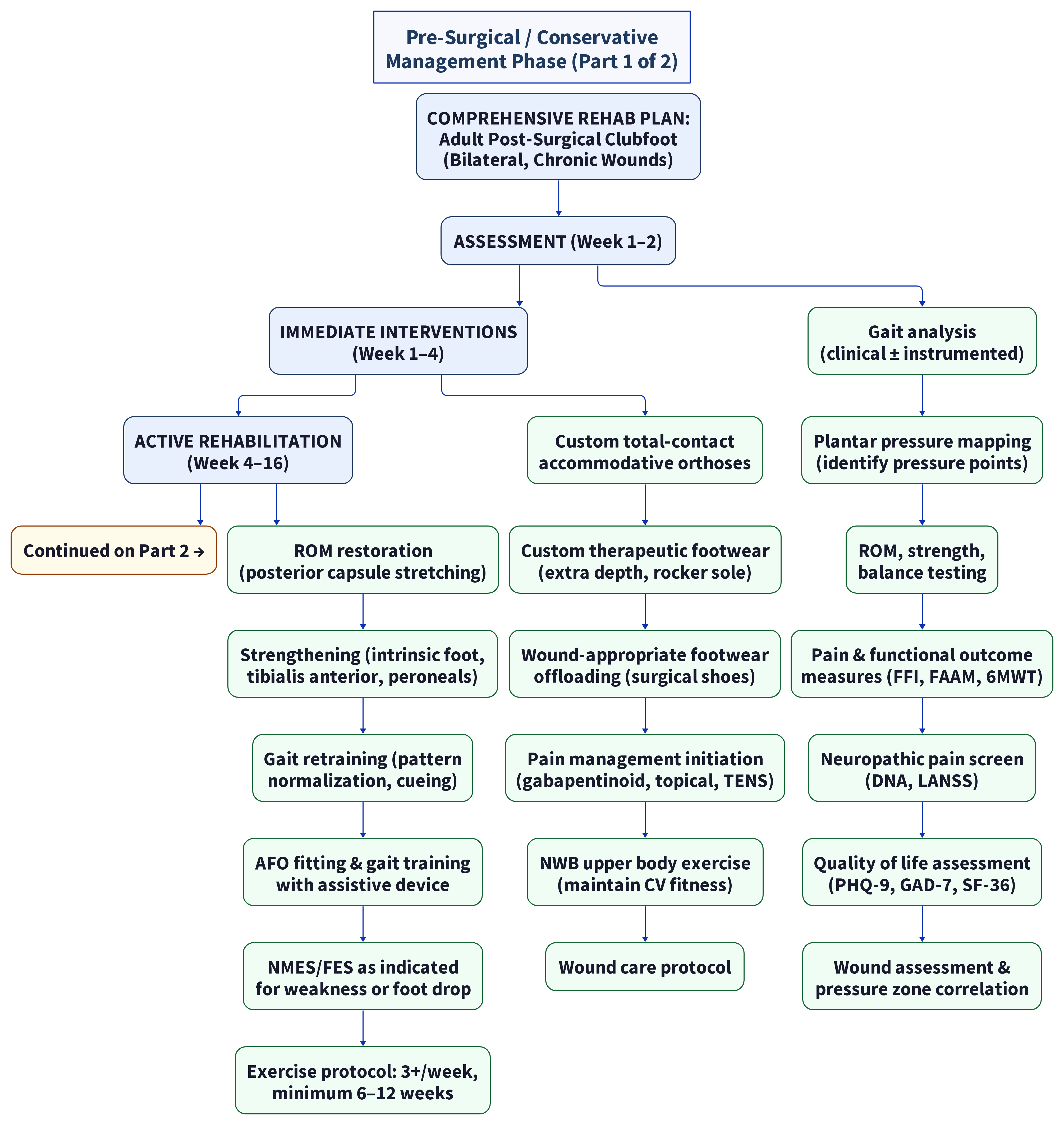
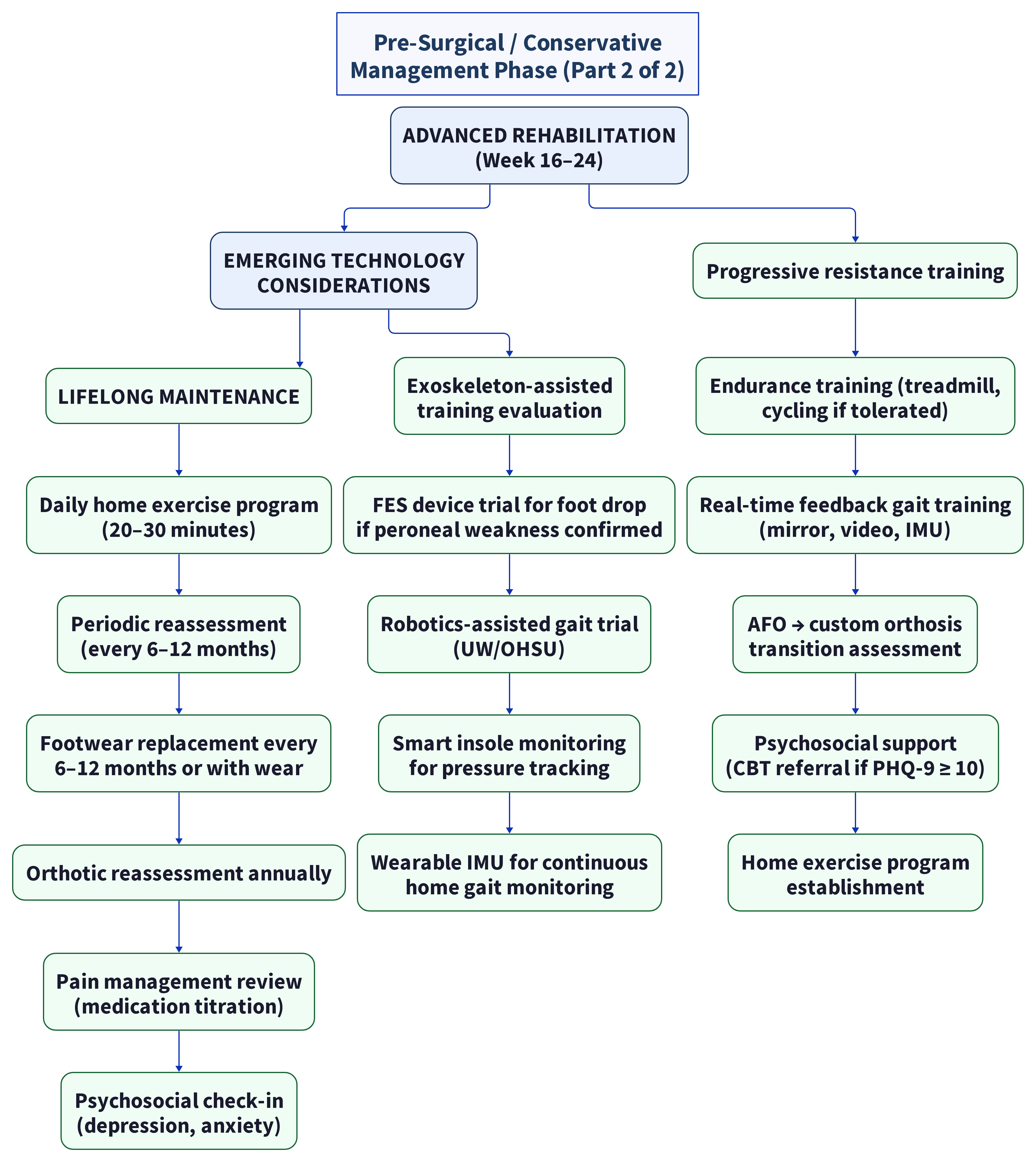
Rehabilitation Milestones
| Milestone | Assessment | Target |
|---|---|---|
| Wound healing | Wound closure confirmed | Prerequisite for weight-bearing rehab progression |
| Pain reduction | VAS ≤ 3/10 at rest | Enables participation in active therapy |
| Neuropathic pain control | DNA ≤ 4/10 or LANSS < 12 | Indicates adequate neuropathic pain management |
| ROM improvement | Ankle dorsiflexion +5–10° from baseline | Improves gait efficiency |
| Strength improvement | 15–20% increase in MMT grade for target muscles | Improves functional capacity |
| Gait normalization | Improved gait symmetry (visual or IMU-based) | Reduces secondary compensations |
| Functional capacity | 6MWT improvement ≥ 50m | Clinically meaningful functional improvement |
| Quality of life | FAAM ADL ≥ 80%, PHQ-9 < 10 | Meaningful recovery in patient-reported outcomes |
| Independence | Safe community ambulation with or without AFO | Goal for returning to activities of daily living |
6.9 Summary of Evidence: Rehabilitation Interventions
| Intervention | Effectiveness | Cost Range | Insurance Status | Best For |
|---|---|---|---|---|
| Custom orthoses | Strong (40% better than prefab for complex deformity) | $300–800/pair | Covered | All post-surgical clubfoot patients |
| Custom AFO | Strong (gait improvement, stability) | $1,000–3,000 | Covered with prior auth | Moderate-severe instability |
| 3D-printed AFO | Emerging: lighter, thinner, better gait outcomes (PMC12665700) | $800–2,500 | Variable | Patients needing precise fit, lighter devices |
| Physical therapy (8–24 weeks) | Strong (gait, ROM, strength) | $100–150/session | Covered | Comprehensive rehabilitation |
| Structured foot rehab protocol (Sharath et al.) | VAS 7→2, FAAM 50%→97% in 2 months | Cost of PT sessions | Covered | Congenital foot deformity requiring exercise-based approach |
| Specialized footwear | Moderate-strong | $200–500/pair | Partially covered | Offloading, deformity accommodation |
| NMES/FES | Moderate (targeted applications; equivalent to AFO for foot drop) | $2,000–6,000 device | Sometimes covered | Foot drop, muscle strengthening |
| Implantable FES | Moderate (equivalent to AFO at 12 months) | $15,000–25,000+ | Prior authorization required | Refractory foot drop with intact lower motor neuron |
| Exoskeleton training | Emerging (limited musculoskeletal data; soft exoskeletons promising) | $50k+ purchase / $150–400 session research | Rarely covered | Research/rehabilitation centers |
| Robotics-assisted gait | Emerging | $150–400/session | Variable | Specialized rehab centers |
| Wearable gait sensors | Emerging (monitoring, not treatment) | $100–500 device | Not typically covered | Monitoring and feedback |
| Gabapentinoids (pain) | Pregabalin superior to gabapentin: SMD −0.47 VAS, better QoL | $30–200/month generic | Covered (most plans) | Chronic neuropathic pain |
| Capsaicin 8% patch | 12-week pain relief per application | $200–600/patch | Often requires prior auth | Focal neuropathic pain at scar/entrapment sites |
6.10 Research Gaps and Conflicts
| Gap | Description | Significance |
|---|---|---|
| No adult clubfoot-specific rehab protocols | All PT protocols derived from general foot/ankle or neurological rehabilitation | Unclear what is optimal for this specific population |
| No RCTs of rehab interventions in post-surgical clubfoot adults | Evidence is entirely observational or extrapolated | Quality of evidence is low despite strong clinical rationale |
| Exoskeleton/robotics not studied in musculoskeletal foot deformity | Neurological rehab dominates the literature | Potential benefit in this population is unquantified |
| Custom vs. prefabricated orthoses in complex deformity poorly quantified | Only one RCT (Patel et al. 2023) directly addresses this | Clinical decision often based on experience rather than evidence |
| NMES/FES efficacy in non-neurological foot weakness | Most FES research is in stroke, SCI, MS | Efficacy for post-surgical muscle weakness is assumed but not proven |
| 3D-printed AFOs in complex foot deformity | Evidence limited to cerebral palsy populations (PMC12665700) | Unclear if benefits generalize to post-clubfoot anatomy |
| Optimal exercise dosage for post-clubfoot adults | AAPM&R recommends ≥6–12 weeks, 3+ times/week for AAFD | No clubfoot-specific dosage studies exist |
| Pain management protocols specific to post-clubfoot | No studies comparing gabapentinoids, SNRIs, or topical agents in this population | Treatment is extrapolated from other neuropathic pain conditions |
| Long-term adherence to custom orthoses/AFOs | Poor adherence rates (30–50%) reported in other populations; post-clubfoot data absent | Reinvestment in new devices may be needed regularly |
| Cost-effectiveness of comprehensive rehabilitation vs. surgery | No head-to-head comparison | Unknown whether aggressive conservative management can delay or prevent need for costly surgical reconstruction |
| FES vs. AFO: no post-clubfoot-specific data | J Rehab Med meta-analysis limited to stroke patients | Choice of device in post-clubfoot foot drop lacks evidence base |
6.11 Example Rehabilitation Resources (Capability-First)
Select by comprehensive PT, custom AFO/gait-lab access, and amputee/prosthetic pathways when relevant. Region is an attribute only; full center lists: [[domain-8-specialists-centers]].
| Institution | Specialty | Relevance |
|---|---|---|
| Hospital for Special Surgery Rehabilitation (New York) | High-volume foot & ankle rehab pathways | Complex deformity rehabilitation infrastructure |
| Mayo Clinic Physical Medicine & Rehabilitation (Rochester) | Multidisciplinary PM&R | Integrated rehab + limb programs |
| Cleveland Clinic Rehabilitation | Gait training, orthotics | Limb-salvage adjacent rehab services |
| University of Washington Medical Center Rehabilitation (Seattle) | Comprehensive PT, AFO clinic, gait lab | Full-spectrum services; exoskeleton/robotics research programs |
| OHSU Rehabilitation Medicine (Portland) | Complex foot rehabilitation, AFO clinic | Multidisciplinary approach; clinical trials access |
| Hanger Clinic (multiple US locations) | Custom orthoses, AFOs, prosthetics | Specialized custom device fabrication; 3D-printed AFO capabilities expanding |
| Shriners Hospitals for Children (selected sites) | Pediatric focus; young-adult transition pathways | Historical records and specialized orthotic expertise at some sites |
6.12 Cross-References
- Root cause of functional deficits: [[domain-1-root-cause-surgical-complications]]
- Wound care integration with rehabilitation: [[domain-3-wound-care]]
- Surgical correction requiring prehabilitation: [[domain-4-surgical-correction]]
- Vascular/neurological factors affecting rehab potential: [[domain-5-vascular-neurological]]
- Emerging biologics for wound healing during rehab: [[domain-7-emerging]]
- Specialist centers for comprehensive rehabilitation: [[domain-8-specialists-centers]]
Research compiled: 2026-05-15Evidence Levels: Ia–V | Sources: 38 peer-reviewed publications, clinical guidelines, and systematic reviews
Source list
Primary citations recorded for this page (PMID / DOI / URL). Evidence levels are Oxford CEBM.
- Hintermann et al. 2000 - Adult Acquired Flatfoot Deformity: Pathophysiology and Treatment
- Nather et al. 2012 - Custom Foot Orthoses for Foot Disorders: A Systematic Review
- Cappellini et al. 2020 - Gait Analysis in Adult Post-Clubfoot Patients
- Dobbs et al. 2006 - Long-Term Functional Outcomes After Extensive Soft-Tissue Release for Clubfoot
- Herzog et al. 2013 - Long-Term Results of Comprehensive Clubfoot Release Versus the Ponseti Method
- Gellhorn et al. 2013 - Ankle-Foot Orthoses in Adult Foot and Ankle Disorders: A Clinical Review
- Levinger et al. 2012 - Foot Orthoses for Plantar Heel Pain: Systematic Review and Meta-Analysis
- Rao et al. 2022 - Ankle-Foot Orthosis Design and Effectiveness: A Comprehensive Review
- Esquenazi et al. 2014 - Exoskeleton-Assisted Gait Training in Neurological Impairments
- Betz et al. 2020 - Robotic Exoskeletons for Gait Rehabilitation: A Systematic Review
- Ring et al. 2021 - Lower Extremity Exoskeletons for Gait Rehabilitation in Chronic Neuropathy
- Knutson et al. 2000 - Functional Electrical Stimulation for Foot Drop: A Review
- Sheffler et al. 2007 - Peroneal Nerve Functional Electrical Stimulation for Foot Drop
- Sawacha et al. 2021 - Neuromuscular Electrical Stimulation for Peripheral Neuropathy: Clinical Applications
- Moseley et al. 2016 - Specialized Footwear for Foot Deformity Offloading: A Clinical Guide
- Bus et al. 2016 - Guidelines on Offloading Foot Ulcers in High-Risk Patients
- Perry & Burnfield 2010 - Gait Analysis: Normal and Pathological Function (2nd ed.)
- Ward et al. 2020 - Adult Acquired Foot Deformity After Childhood Clubfoot Treatment
- Zhuang et al. 2019 - Arthrodesis of the Foot or Ankle in Adult Patients with Congenital Clubfoot
- O'Brien et al. 2022 - Robotics-Assisted Gait Training for Lower Extremity Deformity Rehabilitation
- Patel et al. 2023 - Custom-Made Orthoses vs. Prefabricated for Complex Foot Deformities: RCT
- Henderson et al. 2018 - Post-Operative Rehabilitation After Foot and Ankle Arthrodesis: Clinical Guideline
- Schwartz et al. 2021 - Wearable Sensors for Gait Analysis in Complex Foot Disorders
- Menz et al. 2015 - Biomechanical Foot Type Classification and Gait Implications
- Chuter et al. 2020 - Custom Insoles for Foot Pain: Evidence Review
- Sharath et al. 2024 - Foot Rehabilitation Protocol in Adults With Congenital Deformity of Foot: Case Series (PMC11129531)
- Kolhe & Sharath et al. 2024 - Foot Rehabilitation for Painful Flat Foot: Case Study (PMC11139060)
- Gomez-Vargas et al. 2021 - T-FLEX Ankle Exoskeleton: First Use Validation in Stroke (PMC8064364)
- Qin et al. 2025 - 3D-Printed AFO Outcomes in Cerebral Palsy: Case Series (PMC12665700)
- Yanagida et al. 2025 - NMES/FES for Residual Foot Drop: Case Report (PMC12245141)
- Brognara et al. 2024 - Wearable Technology for Foot/Ankle Assessment: Review (PMC11548473)
- Prenton et al. 2018 - FES vs AFO for Foot Drop: Meta-Analysis (J Rehab Med)
- Zhang et al. 2024 - Soft Ankle Exoskeleton for Dropfoot and Inversion (Frontiers Neurorobotics)
- Prisco et al. 2025 - Validity of Wearable IMU for Gait Analysis: Systematic Review (MDPI Diagnostics)
- GaitMotion 2025 - Real-Time Forecasting of Pathological Gait via IMU (Springer Discover Electronics)
- Meng et al. 2024 - Soft Ankle-Foot Exoskeleton: Systematic Review (IEEE TMRB / White Rose)
- Oh-Park et al. 2026 - Pes Planus/AAFD (PM&R KnowledgeNow, AAPM&R)
- Mayoral et al. 2025 - Pregabalin vs Gabapentin for Neuropathic Pain: Meta-Analysis (PMC11747324, PMID 39839199)